 Movie
Movie Controller

[English] 日本語
 Yorodumi
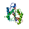
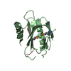
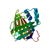
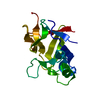
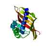
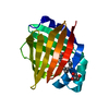
- PDB-2hw2: Crystal structure of Rifampin ADP-ribosyl transferase in complex ... -
+ Open data

- Basic information
| Entry | Database: PDB / ID: 2hw2 | ||||||
|---|---|---|---|---|---|---|---|
| Title | Crystal structure of Rifampin ADP-ribosyl transferase in complex with Rifampin | ||||||
 Components Components | Rifampin ADP-ribosyl transferase | ||||||
 Keywords Keywords | TRANSFERASE / PROTEIN-ANTIBIOTIC COMPLEX / ADP-RIBOSYLATION / RIFAMPIN | ||||||
| Function / homology |  Function and homology information Function and homology information | ||||||
| Biological species |  Mycobacterium smegmatis (bacteria) Mycobacterium smegmatis (bacteria) | ||||||
| Method |  X-RAY DIFFRACTION / SYNCHROTRON / SAD / Resolution: 1.45 Å X-RAY DIFFRACTION / SYNCHROTRON / SAD / Resolution: 1.45 Å | ||||||
 Authors Authors | Baysarowich, J. / Wright, G.D. / Junop, M. | ||||||
 Citation Citation | Journal: Proc.Natl.Acad.Sci.Usa / Year: 2008 Title: Rifamycin antibiotic resistance by ADP-ribosylation: Structure and diversity of Arr. Authors: Baysarowich, J. / Koteva, K. / Hughes, D.W. / Ejim, L. / Griffiths, E. / Zhang, K. / Junop, M. / Wright, G.D. | ||||||
| History |
| ||||||
| Remark 999 | SEQUENCE The difference in sequence is due to a different strain of Mycobacterium smegmatis mc2-155 ...SEQUENCE The difference in sequence is due to a different strain of Mycobacterium smegmatis mc2-155 used for crystallization (Alexander, D., Jones, J., and Liu, J. (2003) Antimicrobial Agents and Chemotherapy, 47, 3208-3213) |
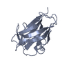
- Structure visualization
| Structure viewer | Molecule: MolmilJmol/JSmol |
|---|
- Downloads & links
-Download
| PDBx/mmCIF format | 2hw2.cif.gz | 84.6 KB | Display | PDBx/mmCIF format |
|---|---|---|---|---|
| PDB format | pdb2hw2.ent.gz | 63.4 KB | Display | PDB format |
| PDBx/mmJSON format | 2hw2.json.gz | Tree view | PDBx/mmJSON format | |
| Others |  Other downloads Other downloads |
-Validation report
| Arichive directory | https://data.pdbj.org/pub/pdb/validation_reports/hw/2hw2ftp://data.pdbj.org/pub/pdb/validation_reports/hw/2hw2 | HTTPS FTP |
|---|
-Related structure data
| Similar structure data |
|---|


-Links
 PDBj
PDBj

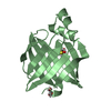
- Assembly
| Deposited unit | 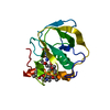
| ||||||||
|---|---|---|---|---|---|---|---|---|---|
| 1 |
| ||||||||
| Unit cell |
| ||||||||
| Components on special symmetry positions |
| ||||||||
| Details | The biological unit is a monomer. |
-Components
| #1: Protein | Mass: 15875.920 Da / Num. of mol.: 1 Source method: isolated from a genetically manipulated source Source: (gene. exp.) Mycobacterium smegmatis (bacteria) / Gene: arr / Plasmid: pET22b / Species (production host): Escherichia coli / Production host: References: UniProt: O67972, UniProt: A0QRS5*PLUS, NAD+ ADP-ribosyltransferase | ||||
|---|---|---|---|---|---|
| #2: Chemical |   Type: peptide linking / Mass: 75.067 Da / Num. of mol.: 2 / Source method: obtained synthetically / Formula: C2H5NO2 Type: peptide linking / Mass: 75.067 Da / Num. of mol.: 2 / Source method: obtained synthetically / Formula: C2H5NO2#3: Chemical | ChemComp-RFP / | 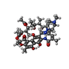  Mass: 822.940 Da / Num. of mol.: 1 / Source method: obtained synthetically / Formula: C43H58N4O12 / Comment: antibiotic*YM Mass: 822.940 Da / Num. of mol.: 1 / Source method: obtained synthetically / Formula: C43H58N4O12 / Comment: antibiotic*YM#4: Water | ChemComp-HOH / |  Mass: 18.015 Da / Num. of mol.: 254 / Source method: isolated from a natural source / Formula: H2O Mass: 18.015 Da / Num. of mol.: 254 / Source method: isolated from a natural source / Formula: H2O |
-Experimental details
-Experiment
| Experiment | Method: X-RAY DIFFRACTION / Number of used crystals: 1 |
|---|
- Sample preparation
| Crystal | Density Matthews: 2.42 Å3/Da / Density % sol: 49.07 % |
|---|---|
| Crystal grow | Temperature: 298 K / Method: vapor diffusion, sitting drop / pH: 8.2 Details: 30% MPD, 5% PEG 8000, 0.005M magesium chloride, 0.001M rifampin, 0.03M glycylglycine, 0.05M tris, pH 8.2, VAPOR DIFFUSION, SITTING DROP, temperature 298.0K |
-Data collection
| Diffraction | Mean temperature: 100 K |
|---|---|
| Diffraction source | Source: SYNCHROTRON / Site: NSLS  / Beamline: X8C / Wavelength: 1.1 Å / Beamline: X8C / Wavelength: 1.1 Å |
| Detector | Type: ADSC QUANTUM 4 / Detector: CCD / Date: Jun 11, 2006 |
| Radiation | Monochromator: Silicon 111 / Protocol: SINGLE WAVELENGTH / Monochromatic (M) / Laue (L): M / Scattering type: x-ray |
| Radiation wavelength | Wavelength: 1.1 Å / Relative weight: 1 |
| Reflection | Resolution: 1.45→30.18 Å / Num. all: 26649 / Num. obs: 26649 / % possible obs: 98 % / Observed criterion σ(F): 1 / Observed criterion σ(I): 1 / Redundancy: 4.5 % / Rmerge(I) obs: 0.058 / Net I/σ(I): 20.6 |
| Reflection shell | Resolution: 1.45→1.5 Å / Redundancy: 4 % / Rmerge(I) obs: 0.544 / Mean I/σ(I) obs: 2.7 / Num. unique all: 2578 / % possible all: 95.9 |
- Processing
| Software |
| ||||||||||||||||||||||||||||||||||||||||||||||||||||||||||||||||||||||||||||||||||||||||||||||||||||
|---|---|---|---|---|---|---|---|---|---|---|---|---|---|---|---|---|---|---|---|---|---|---|---|---|---|---|---|---|---|---|---|---|---|---|---|---|---|---|---|---|---|---|---|---|---|---|---|---|---|---|---|---|---|---|---|---|---|---|---|---|---|---|---|---|---|---|---|---|---|---|---|---|---|---|---|---|---|---|---|---|---|---|---|---|---|---|---|---|---|---|---|---|---|---|---|---|---|---|---|---|---|
| Refinement | Method to determine structure: SAD / Resolution: 1.45→30.18 Å / Cor.coef. Fo:Fc: 0.978 / Cor.coef. Fo:Fc free: 0.962 / SU B: 1.501 / SU ML: 0.027 / Cross valid method: THROUGHOUT / σ(F): 1 / ESU R: 0.068 / ESU R Free: 0.066 / Stereochemistry target values: MAXIMUM LIKELIHOOD
| ||||||||||||||||||||||||||||||||||||||||||||||||||||||||||||||||||||||||||||||||||||||||||||||||||||
| Solvent computation | Ion probe radii: 0.8 Å / Shrinkage radii: 0.8 Å / VDW probe radii: 1.4 Å / Solvent model: BABINET MODEL WITH MASK | ||||||||||||||||||||||||||||||||||||||||||||||||||||||||||||||||||||||||||||||||||||||||||||||||||||
| Displacement parameters | Biso mean: 29.554 Å2
| ||||||||||||||||||||||||||||||||||||||||||||||||||||||||||||||||||||||||||||||||||||||||||||||||||||
| Refinement step | Cycle: LAST / Resolution: 1.45→30.18 Å
| ||||||||||||||||||||||||||||||||||||||||||||||||||||||||||||||||||||||||||||||||||||||||||||||||||||
| Refine LS restraints |
| ||||||||||||||||||||||||||||||||||||||||||||||||||||||||||||||||||||||||||||||||||||||||||||||||||||
| LS refinement shell | Resolution: 1.45→1.486 Å / Total num. of bins used: 20
| ||||||||||||||||||||||||||||||||||||||||||||||||||||||||||||||||||||||||||||||||||||||||||||||||||||
| Refinement TLS params. | Method: refined / Refine-ID: X-RAY DIFFRACTION
| ||||||||||||||||||||||||||||||||||||||||||||||||||||||||||||||||||||||||||||||||||||||||||||||||||||
| Refinement TLS group |
|
