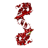
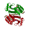
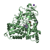
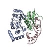
BIOMOLECULE: 1 THIS ENTRY CONTAINS THE CRYSTALLOGRAPHIC ASYMMETRIC UNIT WHICH CONSISTS OF 2 CHAINS. ... BIOMOLECULE: 1 THIS ENTRY CONTAINS THE CRYSTALLOGRAPHIC ASYMMETRIC UNIT WHICH CONSISTS OF 2 CHAINS. SEE REMARK 350 FOR INFORMATION ON GENERATING THE BIOLOGICAL MOLECULE. THE BIOLOGICAL UNIT IS A DIMER FORMED BY DISULFIDE BOND BETWEEN CYSTEINES 72. THE DIMER IS PRESENT ENTIRELY IN THE ASYMMETRIC UNIT AND DOES NOT HAVE A CRYSTALLOGRAPHIC SYMMETRY.
Mass: 18.015 Da / Num. of mol.: 99 / Source method: isolated from a natural source / Formula: H2O
Has protein modification
Y
-
Experimental details
-
Experiment
Experiment
Method: X-RAY DIFFRACTION / Number of used crystals: 1
-
Sample preparation
Crystal
Density Matthews: 3.17 Å3/Da / Density % sol: 61.17 % Description: Terminal residues 163 and 164 for T4 lysozyme have poor electron density. This should be taken into account when using this structure as a model.
Crystal grow
Temperature: 298 K / Method: vapor diffusion, hanging drop / pH: 7 Details: 0.2 M tri-Lithium Citrate, 20% PEG 3350, pH 7.0, VAPOR DIFFUSION, HANGING DROP, temperature 298K
-
Data collection
Diffraction
Mean temperature: 200 K
Diffraction source
Source: SYNCHROTRON / Site: ALS / Beamline: 8.2.2 / Wavelength: 1 Å
Resolution: 2.35→52.6 Å / Cor.coef. Fo:Fc: 0.891 / Cor.coef. Fo:Fc free: 0.839 / SU B: 14.465 / SU ML: 0.184 / Cross valid method: THROUGHOUT / σ(F): 2 / ESU R: 0.341 / ESU R Free: 0.278 / Stereochemistry target values: MAXIMUM LIKELIHOOD Details: HYDROGENS HAVE BEEN ADDED IN THE RIDING POSITIONS. DATA WAS COLLECTED TO 2.10 ANGSTROM RESOLUTION. STRUCTURE WAS REFINED TO 2.35 ANGSTROM RESOLUTION.
Rfactor
Num. reflection
% reflection
Selection details
Rfree
0.30355
993
5.1 %
RANDOM
Rwork
0.23939
-
-
-
obs
0.24277
18576
99.67 %
-
all
-
19569
-
-
Solvent computation
Ion probe radii: 0.8 Å / Shrinkage radii: 0.8 Å / VDW probe radii: 1.4 Å / Solvent model: MASK
Displacement parameters
Biso mean: 21.3 Å2
Baniso -1
Baniso -2
Baniso -3
1-
1.11 Å2
0 Å2
0.63 Å2
2-
-
-1.86 Å2
0 Å2
3-
-
-
1.14 Å2
Refinement step
Cycle: LAST / Resolution: 2.35→52.6 Å
Protein
Nucleic acid
Ligand
Solvent
Total
Num. atoms
2612
0
8
99
2719
Refine LS restraints
Refine-ID
Type
Dev ideal
Dev ideal target
Number
X-RAY DIFFRACTION
r_bond_refined_d
0.026
0.022
2660
X-RAY DIFFRACTION
r_bond_other_d
X-RAY DIFFRACTION
r_angle_refined_deg
2.08
1.956
3583
X-RAY DIFFRACTION
r_angle_other_deg
X-RAY DIFFRACTION
r_dihedral_angle_1_deg
7.128
5
326
X-RAY DIFFRACTION
r_dihedral_angle_2_deg
34.825
23.387
124
X-RAY DIFFRACTION
r_dihedral_angle_3_deg
18.106
15
496
X-RAY DIFFRACTION
r_dihedral_angle_4_deg
16.462
15
26
X-RAY DIFFRACTION
r_chiral_restr
0.133
0.2
400
X-RAY DIFFRACTION
r_gen_planes_refined
0.009
0.02
1968
X-RAY DIFFRACTION
r_gen_planes_other
X-RAY DIFFRACTION
r_nbd_refined
0.226
0.2
1285
X-RAY DIFFRACTION
r_nbd_other
X-RAY DIFFRACTION
r_nbtor_refined
0.303
0.2
1812
X-RAY DIFFRACTION
r_nbtor_other
X-RAY DIFFRACTION
r_xyhbond_nbd_refined
0.197
0.2
154
X-RAY DIFFRACTION
r_xyhbond_nbd_other
X-RAY DIFFRACTION
r_metal_ion_refined
X-RAY DIFFRACTION
r_metal_ion_other
X-RAY DIFFRACTION
r_symmetry_vdw_refined
0.297
0.2
31
X-RAY DIFFRACTION
r_symmetry_vdw_other
X-RAY DIFFRACTION
r_symmetry_hbond_refined
0.193
0.2
11
X-RAY DIFFRACTION
r_symmetry_hbond_other
X-RAY DIFFRACTION
r_symmetry_metal_ion_refined
X-RAY DIFFRACTION
r_symmetry_metal_ion_other
X-RAY DIFFRACTION
r_mcbond_it
1.478
1.5
1721
X-RAY DIFFRACTION
r_mcbond_other
X-RAY DIFFRACTION
r_mcangle_it
1.954
2
2606
X-RAY DIFFRACTION
r_scbond_it
3.514
3
1149
X-RAY DIFFRACTION
r_scangle_it
5.075
4.5
977
X-RAY DIFFRACTION
r_rigid_bond_restr
X-RAY DIFFRACTION
r_sphericity_free
X-RAY DIFFRACTION
r_sphericity_bonded
LS refinement shell
Resolution: 2.35→2.411 Å / Total num. of bins used: 20
Rfactor
Num. reflection
% reflection
Rfree
0.334
76
-
Rwork
0.215
1351
-
obs
-
-
99.44 %
+
About Yorodumi
-
News
-
Feb 9, 2022. New format data for meta-information of EMDB entries
New format data for meta-information of EMDB entries
Version 3 of the EMDB header file is now the official format.
The previous official version 1.9 will be removed from the archive.
In the structure databanks used in Yorodumi, some data are registered as the other names, "COVID-19 virus" and "2019-nCoV". Here are the details of the virus and the list of structure data.
Jan 31, 2019. EMDB accession codes are about to change! (news from PDBe EMDB page)
EMDB accession codes are about to change! (news from PDBe EMDB page)
The allocation of 4 digits for EMDB accession codes will soon come to an end. Whilst these codes will remain in use, new EMDB accession codes will include an additional digit and will expand incrementally as the available range of codes is exhausted. The current 4-digit format prefixed with “EMD-” (i.e. EMD-XXXX) will advance to a 5-digit format (i.e. EMD-XXXXX), and so on. It is currently estimated that the 4-digit codes will be depleted around Spring 2019, at which point the 5-digit format will come into force.
The EM Navigator/Yorodumi systems omit the EMD- prefix.
Related info.:Q: What is EMD? / ID/Accession-code notation in Yorodumi/EM Navigator
Yorodumi is a browser for structure data from EMDB, PDB, SASBDB, etc.
This page is also the successor to EM Navigator detail page, and also detail information page/front-end page for Omokage search.
The word "yorodu" (or yorozu) is an old Japanese word meaning "ten thousand". "mi" (miru) is to see.
Related info.:EMDB / PDB / SASBDB / Comparison of 3 databanks / Yorodumi Search / Aug 31, 2016. New EM Navigator & Yorodumi / Yorodumi Papers / Jmol/JSmol / Function and homology information / Changes in new EM Navigator and Yorodumi
 Movie
Movie Controller


 Open data
 Basic information
 Components
 Keywords
 Function and homology information
 Enterobacteria phage T4 (virus)
 X-RAY DIFFRACTION /
  Authors
 Citation
 Structure visualization
 Downloads & links
 Other downloads
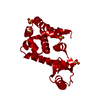


 PDBj
PDBj


 Assembly


 Mass: 122.143 Da / Num. of mol.: 1 / Source method: obtained synthetically / Formula: C4H12NO3 / Comment: pH buffer*YM
 Mass: 18.015 Da / Num. of mol.: 99 / Source method: isolated from a natural source / Formula: H2O
 Sample preparation
 / Beamline: 8.2.2 / Wavelength: 1 Å
 Processing