- PDB-2hth: Structural basis for ubiquitin recognition by the human EAP45/ESC... -
+
Open data
ID or keywords:
Loading...
-
Basic information
Entry
Database: PDB / ID: 2hth
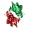
Title
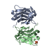
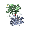
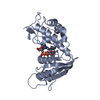
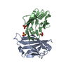
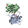
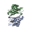
Structural basis for ubiquitin recognition by the human EAP45/ESCRT-II GLUE domain
Components
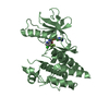
Ubiquitin
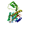
Vacuolar protein sorting protein 36
Keywords
PROTEIN TRANSPORT / GLUE domain / PH domain / Protein sorting / viral budding / Ubiquitin complex
Function / homology
Function and homology information
ESCRT II complex / : / : / protein modification process => GO:0036211 / : / protein transport to vacuole involved in ubiquitin-dependent protein catabolic process via the multivesicular body sorting pathway / membrane fission / multivesicular body assembly / phosphatidylinositol-3-phosphate binding / Peptide chain elongation ...ESCRT II complex / : / : / protein modification process => GO:0036211 / : / protein transport to vacuole involved in ubiquitin-dependent protein catabolic process via the multivesicular body sorting pathway / membrane fission / multivesicular body assembly / phosphatidylinositol-3-phosphate binding / Peptide chain elongation / Selenocysteine synthesis / Formation of a pool of free 40S subunits / Eukaryotic Translation Termination / SRP-dependent cotranslational protein targeting to membrane / Response of EIF2AK4 (GCN2) to amino acid deficiency / Viral mRNA Translation / Nonsense Mediated Decay (NMD) independent of the Exon Junction Complex (EJC) / GTP hydrolysis and joining of the 60S ribosomal subunit / L13a-mediated translational silencing of Ceruloplasmin expression / Major pathway of rRNA processing in the nucleolus and cytosol / Nonsense Mediated Decay (NMD) enhanced by the Exon Junction Complex (EJC) / Maturation of protein E / Maturation of protein E / ER Quality Control Compartment (ERQC) / Myoclonic epilepsy of Lafora / FLT3 signaling by CBL mutants / IRAK2 mediated activation of TAK1 complex / Alpha-protein kinase 1 signaling pathway / Glycogen synthesis / IRAK1 recruits IKK complex / IRAK1 recruits IKK complex upon TLR7/8 or 9 stimulation / Prevention of phagosomal-lysosomal fusion / Endosomal Sorting Complex Required For Transport (ESCRT) / Membrane binding and targetting of GAG proteins / Regulation of TBK1, IKKε (IKBKE)-mediated activation of IRF3, IRF7 / Negative regulation of FLT3 / PTK6 Regulates RTKs and Their Effectors AKT1 and DOK1 / Regulation of TBK1, IKKε-mediated activation of IRF3, IRF7 upon TLR3 ligation / IRAK2 mediated activation of TAK1 complex upon TLR7/8 or 9 stimulation / Constitutive Signaling by NOTCH1 HD Domain Mutants / NOTCH2 Activation and Transmission of Signal to the Nucleus / TICAM1,TRAF6-dependent induction of TAK1 complex / TICAM1-dependent activation of IRF3/IRF7 / APC/C:Cdc20 mediated degradation of Cyclin B / Downregulation of ERBB4 signaling / APC-Cdc20 mediated degradation of Nek2A / Regulation of FZD by ubiquitination / p75NTR recruits signalling complexes / cytosolic ribosome / InlA-mediated entry of Listeria monocytogenes into host cells / TRAF6 mediated IRF7 activation in TLR7/8 or 9 signaling / NF-kB is activated and signals survival / TRAF6-mediated induction of TAK1 complex within TLR4 complex / Regulation of pyruvate metabolism / Pexophagy / Downregulation of ERBB2:ERBB3 signaling / Regulation of innate immune responses to cytosolic DNA / NRIF signals cell death from the nucleus / Regulation of PTEN localization / VLDLR internalisation and degradation / Activated NOTCH1 Transmits Signal to the Nucleus / Synthesis of active ubiquitin: roles of E1 and E2 enzymes / Translesion synthesis by REV1 / TICAM1, RIP1-mediated IKK complex recruitment / HCMV Late Events / Regulation of BACH1 activity / Translesion synthesis by POLK / JNK (c-Jun kinases) phosphorylation and activation mediated by activated human TAK1 / InlB-mediated entry of Listeria monocytogenes into host cell / MAP3K8 (TPL2)-dependent MAPK1/3 activation / Activation of IRF3, IRF7 mediated by TBK1, IKKε (IKBKE) / Downregulation of TGF-beta receptor signaling / Translesion synthesis by POLI / Josephin domain DUBs / Gap-filling DNA repair synthesis and ligation in GG-NER / IKK complex recruitment mediated by RIP1 / PINK1-PRKN Mediated Mitophagy / TGF-beta receptor signaling in EMT (epithelial to mesenchymal transition) / TNFR1-induced NF-kappa-B signaling pathway / macroautophagy / Regulation of activated PAK-2p34 by proteasome mediated degradation / TCF dependent signaling in response to WNT / Regulation of NF-kappa B signaling / activated TAK1 mediates p38 MAPK activation / ubiquitin binding / Autodegradation of Cdh1 by Cdh1:APC/C / APC/C:Cdc20 mediated degradation of Securin / NOTCH3 Activation and Transmission of Signal to the Nucleus / Regulation of signaling by CBL / Negative regulators of DDX58/IFIH1 signaling / N-glycan trimming in the ER and Calnexin/Calreticulin cycle / Asymmetric localization of PCP proteins / Fanconi Anemia Pathway / Negative regulation of FGFR3 signaling / Ubiquitin-dependent degradation of Cyclin D / Peroxisomal protein import / Deactivation of the beta-catenin transactivating complex / SCF-beta-TrCP mediated degradation of Emi1 / NIK-->noncanonical NF-kB signaling / Stabilization of p53 Similarity search - Function
Vacuolar protein sorting protein 36, GLUE domain / Vacuolar protein sorting protein 36 / Snf8/Vps36 family / EAP30/Vps36 family / Vacuolar protein sorting protein 36 Vps36 / GLUE domain profile. / Pleckstrin-homology domain (PH domain)/Phosphotyrosine-binding domain (PTB) / PH-domain like / Ribosomal L40e family / Ribosomal_L40e ...Vacuolar protein sorting protein 36, GLUE domain / Vacuolar protein sorting protein 36 / Snf8/Vps36 family / EAP30/Vps36 family / Vacuolar protein sorting protein 36 Vps36 / GLUE domain profile. / Pleckstrin-homology domain (PH domain)/Phosphotyrosine-binding domain (PTB) / PH-domain like / Ribosomal L40e family / Ribosomal_L40e / Ribosomal protein L40e / Ribosomal protein L40e superfamily / Phosphatidylinositol 3-kinase Catalytic Subunit; Chain A, domain 1 / Ubiquitin-like (UB roll) / : / Ubiquitin domain signature. / Ubiquitin conserved site / Ubiquitin domain / PH-like domain superfamily / Ubiquitin family / Ubiquitin homologues / Ubiquitin domain profile. / Ubiquitin-like domain / Roll / Ubiquitin-like domain superfamily / Winged helix DNA-binding domain superfamily / Winged helix-like DNA-binding domain superfamily / Roll / Mainly Beta / Alpha Beta Similarity search - Domain/homology
Polyubiquitin-C / Ubiquitin-60S ribosomal protein L40 / Vacuolar protein-sorting-associated protein 36 Similarity search - Component
Biological species
Homo sapiens (human)
Method
X-RAY DIFFRACTION / SYNCHROTRON / SAD / Resolution: 2.7 Å
Monochromator: Si 111 CHANNEL / Protocol: SINGLE WAVELENGTH / Monochromatic (M) / Laue (L): M / Scattering type: x-ray
Radiation wavelength
ID
Wavelength (Å)
Relative weight
1
1.1
1
2
0.9791
1
Reflection
Av σ(I) over netI: 8.1 / Number: 187787 / Rmerge(I) obs: 0.123 / Χ2: 1.01 / D res high: 3.1 Å / D res low: 50 Å / Num. obs: 12326 / % possible obs: 100
Diffraction reflection shell
Highest resolution (Å)
Lowest resolution (Å)
% possible obs (%)
ID
Rmerge(I) obs
Chi squared
6.67
50
99.9
1
0.085
1.023
5.3
6.67
99.9
1
0.102
0.879
4.63
5.3
100
1
0.105
1.065
4.21
4.63
100
1
0.111
1.085
3.91
4.21
100
1
0.139
1.392
3.68
3.91
100
1
0.152
1.408
3.49
3.68
100
1
0.205
1.072
3.34
3.49
100
1
0.254
0.876
3.21
3.34
100
1
0.376
0.685
3.1
3.21
100
1
0.53
0.586
Reflection
Resolution: 2.7→50 Å / Num. obs: 8264 / % possible obs: 97.7 % / Observed criterion σ(F): 0 / Rmerge(I) obs: 0.109 / Net I/σ(I): 11
Method to determine structure: SAD / Resolution: 2.7→37.32 Å / Cor.coef. Fo:Fc: 0.916 / Cor.coef. Fo:Fc free: 0.873 / SU B: 13.718 / SU ML: 0.28 / Cross valid method: THROUGHOUT / σ(F): 0 / ESU R: 0.626 / ESU R Free: 0.355 / Stereochemistry target values: MAXIMUM LIKELIHOOD Details: HYDROGENS HAVE BEEN ADDED IN THE RIDING POSITIONS. LOOP RESIDUES 32-36, 53-57, 74-78 AND 90-103 ARE SET AT ZERO OCCUPANCY.
Rfactor
Num. reflection
% reflection
Selection details
Rfree
0.28872
810
9.8 %
RANDOM
Rwork
0.24775
-
-
-
obs
0.25184
7426
97.73 %
-
Solvent computation
Ion probe radii: 0.8 Å / Shrinkage radii: 0.8 Å / VDW probe radii: 1.2 Å / Solvent model: MASK
Displacement parameters
Biso mean: 56.345 Å2
Baniso -1
Baniso -2
Baniso -3
1-
-1.73 Å2
0 Å2
0 Å2
2-
-
-1.73 Å2
0 Å2
3-
-
-
3.45 Å2
Refinement step
Cycle: LAST / Resolution: 2.7→37.32 Å
Protein
Nucleic acid
Ligand
Solvent
Total
Num. atoms
1640
0
0
0
1640
Refine LS restraints
Refine-ID
Type
Dev ideal
Dev ideal target
Number
X-RAY DIFFRACTION
r_bond_refined_d
0.018
0.022
1436
X-RAY DIFFRACTION
r_bond_other_d
X-RAY DIFFRACTION
r_angle_refined_deg
1.681
1.953
1930
X-RAY DIFFRACTION
r_angle_other_deg
X-RAY DIFFRACTION
r_dihedral_angle_1_deg
7.251
5
167
X-RAY DIFFRACTION
r_dihedral_angle_2_deg
32.972
23.913
69
X-RAY DIFFRACTION
r_dihedral_angle_3_deg
15.795
15
280
X-RAY DIFFRACTION
r_dihedral_angle_4_deg
14
15
12
X-RAY DIFFRACTION
r_chiral_restr
0.107
0.2
224
X-RAY DIFFRACTION
r_gen_planes_refined
0.005
0.02
1038
X-RAY DIFFRACTION
r_gen_planes_other
X-RAY DIFFRACTION
r_nbd_refined
0.201
0.2
518
X-RAY DIFFRACTION
r_nbd_other
X-RAY DIFFRACTION
r_nbtor_refined
0.317
0.2
937
X-RAY DIFFRACTION
r_nbtor_other
X-RAY DIFFRACTION
r_xyhbond_nbd_refined
0.148
0.2
33
X-RAY DIFFRACTION
r_xyhbond_nbd_other
X-RAY DIFFRACTION
r_metal_ion_refined
X-RAY DIFFRACTION
r_metal_ion_other
X-RAY DIFFRACTION
r_symmetry_vdw_refined
0.199
0.2
12
X-RAY DIFFRACTION
r_symmetry_vdw_other
X-RAY DIFFRACTION
r_symmetry_hbond_refined
X-RAY DIFFRACTION
r_symmetry_hbond_other
X-RAY DIFFRACTION
r_symmetry_metal_ion_refined
X-RAY DIFFRACTION
r_symmetry_metal_ion_other
X-RAY DIFFRACTION
r_mcbond_it
1.032
1.5
877
X-RAY DIFFRACTION
r_mcbond_other
X-RAY DIFFRACTION
r_mcangle_it
1.814
2
1385
X-RAY DIFFRACTION
r_scbond_it
2.333
3
601
X-RAY DIFFRACTION
r_scangle_it
3.68
4.5
545
X-RAY DIFFRACTION
r_rigid_bond_restr
X-RAY DIFFRACTION
r_sphericity_free
X-RAY DIFFRACTION
r_sphericity_bonded
LS refinement shell
Resolution: 2.7→2.77 Å / Total num. of bins used: 20
Rfactor
Num. reflection
% reflection
Rfree
0.37
49
-
Rwork
0.367
440
-
obs
-
-
79.64 %
Refinement TLS params.
Method: refined / Refine-ID: X-RAY DIFFRACTION
ID
L11 (°2)
L12 (°2)
L13 (°2)
L22 (°2)
L23 (°2)
L33 (°2)
S11 (Å °)
S12 (Å °)
S13 (Å °)
S21 (Å °)
S22 (Å °)
S23 (Å °)
S31 (Å °)
S32 (Å °)
S33 (Å °)
T11 (Å2)
T12 (Å2)
T13 (Å2)
T22 (Å2)
T23 (Å2)
T33 (Å2)
Origin x (Å)
Origin y (Å)
Origin z (Å)
1
19.634
-16.9771
17.2776
48.4984
-33.2837
31.7127
0.4256
1.1251
0.3466
-2.0371
0.1549
0.0719
0.5117
-0.5583
-0.5804
0.1442
-0.0561
0.1675
0.3623
0
0.6009
-5.3993
-25.0366
-25.8532
2
11.1428
3.0065
1.7129
23.55
-2.6671
7.9247
-0.0635
0.7222
1.6764
-0.2905
0.352
0.4973
-0.8265
-0.4311
-0.2885
0.1873
0.0437
0.1834
0.3439
0.0607
0.638
-4.9871
-18.8278
-22.1207
3
10.611
-4.9379
-2.2412
10.8899
-6.0097
12.4347
-0.6711
-0.5165
0.7323
1.005
0.958
0.2475
-0.6627
-0.6338
-0.2869
0.1711
-0.0085
0.1243
0.1263
0.0068
0.6151
-4.5547
-22.1567
-14.1221
4
14.7253
-12.0584
-12.7211
16.9704
8.7893
54.8173
-0.3704
-1.2615
1.5901
0.6208
0.9662
-0.1425
-0.9035
2.3249
-0.5958
0.1854
-0.1288
-0.0359
0.386
-0.0789
0.8471
4.719
-24.0905
-16.9787
5
19.7679
-0.8298
4.0061
14.4189
-3.1264
1.4202
-0.5057
1.2566
1.0785
-0.8916
-0.206
-0.7966
-0.7334
1.0542
0.7118
0.3623
-0.1263
0.1596
0.3249
0.155
0.7623
6.729
-17.3533
-25.1469
6
9.48
-4.444
1.2532
11.2689
-5.8792
7.6055
0.3157
1.2911
0.7637
-0.7296
-0.5377
-0.3306
-0.3848
0.3145
0.222
0.1396
-0.0147
0.1327
0.2709
0.0627
0.5364
1.0983
-20.9967
-26.2825
7
5.9458
-0.1076
-1.8299
6.8719
4.2756
11.2208
-0.1844
-0.1297
-0.5964
0.3415
-0.3363
0.2974
0.4668
-0.4607
0.5207
0.2069
-0.0006
0.0157
0.1302
0.0043
0.6369
18.3435
-43.9012
-33.3588
8
7.9222
-0.0113
-1.2208
8.6676
7.6621
6.9583
-0.5329
-0.4755
-0.7273
1.1487
0.5099
-0.1685
0.9604
-0.2136
0.0229
0.2665
0.0373
-0.0031
0.1906
0.0089
0.6573
18.3896
-42.119
-29.7728
9
4.0699
-0.2853
-0.7193
6.3016
4.4173
5.3232
-0.1245
-0.4479
-0.5231
0.4937
0.0356
-0.1142
0.766
0.1438
0.0889
0.1617
-0.0435
0.0765
0.2586
0.0714
0.5299
15.7534
-39.4118
-27.1543
10
13.7454
-9.2369
-4.3611
7.8278
2.9229
9.6719
0.348
0.1735
-1.1805
0.0254
-0.2911
0.7587
0.3725
-0.9308
-0.0569
0.2663
-0.059
0.0605
0.2199
0.0465
0.6263
11.9839
-37.392
-22.1819
11
12.7785
3.2136
-1.0869
20.1873
-2.2771
2.7771
-0.3526
-0.6829
-0.2631
0.9876
0.0945
-1.1637
0.1107
0.5612
0.2581
0.1396
0.0657
-0.0364
0.2017
-0.0131
0.4271
25.243
-33.6986
-26.2715
12
29.7743
-4.2892
8.2628
8.3266
-12.7165
19.5273
-0.6613
0.7767
0.1505
-1.1114
0.7561
0.9383
0.6111
-0.4472
-0.0948
0.1483
-0.0171
-0.1132
0.1884
0.0252
0.6013
14.3316
-29.3624
-36.305
Refinement TLS group
ID
Refine-ID
Refine TLS-ID
Auth asym-ID
Label asym-ID
Auth seq-ID
Label seq-ID
1
X-RAY DIFFRACTION
1
A
A
1 - 8
1 - 8
2
X-RAY DIFFRACTION
2
A
A
9 - 26
9 - 26
3
X-RAY DIFFRACTION
3
A
A
27 - 38
27 - 38
4
X-RAY DIFFRACTION
4
A
A
39 - 44
39 - 44
5
X-RAY DIFFRACTION
5
A
A
45 - 55
45 - 55
6
X-RAY DIFFRACTION
6
A
A
56 - 73
56 - 73
7
X-RAY DIFFRACTION
7
B
B
5 - 29
7 - 31
8
X-RAY DIFFRACTION
8
B
B
30 - 47
32 - 49
9
X-RAY DIFFRACTION
9
B
B
48 - 83
50 - 85
10
X-RAY DIFFRACTION
10
B
B
84 - 112
86 - 114
11
X-RAY DIFFRACTION
11
B
B
113 - 124
115 - 126
12
X-RAY DIFFRACTION
12
B
B
125 - 131
127 - 133
+
About Yorodumi
-
News
-
Feb 9, 2022. New format data for meta-information of EMDB entries
New format data for meta-information of EMDB entries
Version 3 of the EMDB header file is now the official format.
The previous official version 1.9 will be removed from the archive.
In the structure databanks used in Yorodumi, some data are registered as the other names, "COVID-19 virus" and "2019-nCoV". Here are the details of the virus and the list of structure data.
Jan 31, 2019. EMDB accession codes are about to change! (news from PDBe EMDB page)
EMDB accession codes are about to change! (news from PDBe EMDB page)
The allocation of 4 digits for EMDB accession codes will soon come to an end. Whilst these codes will remain in use, new EMDB accession codes will include an additional digit and will expand incrementally as the available range of codes is exhausted. The current 4-digit format prefixed with “EMD-” (i.e. EMD-XXXX) will advance to a 5-digit format (i.e. EMD-XXXXX), and so on. It is currently estimated that the 4-digit codes will be depleted around Spring 2019, at which point the 5-digit format will come into force.
The EM Navigator/Yorodumi systems omit the EMD- prefix.
Related info.:Q: What is EMD? / ID/Accession-code notation in Yorodumi/EM Navigator
Yorodumi is a browser for structure data from EMDB, PDB, SASBDB, etc.
This page is also the successor to EM Navigator detail page, and also detail information page/front-end page for Omokage search.
The word "yorodu" (or yorozu) is an old Japanese word meaning "ten thousand". "mi" (miru) is to see.
Related info.:EMDB / PDB / SASBDB / Comparison of 3 databanks / Yorodumi Search / Aug 31, 2016. New EM Navigator & Yorodumi / Yorodumi Papers / Jmol/JSmol / Function and homology information / Changes in new EM Navigator and Yorodumi
 Movie
Movie Controller

 Yorodumi
 Open data
 Basic information
 Components
 Keywords
 Function and homology information
 Homo sapiens (human)
 X-RAY DIFFRACTION /
  Authors
 Citation
 Structure visualization
 Downloads & links
 Other downloads

 PDBj
PDBj


 Assembly
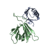

 Sample preparation
 / Beamline: X29A / Wavelength: 1.10000, 0.97910
 Processing