 Movie
Movie Controller

[English] 日本語
 Yorodumi
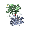
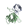
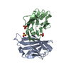
- PDB-4w68: Cytoplasmically Produced Homodimeric Single Domain Antibody (sdAb... -
+ Open data

- Basic information
| Entry | Database: PDB / ID: 4w68 | ||||||
|---|---|---|---|---|---|---|---|
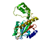
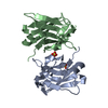
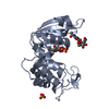
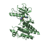
| Title | Cytoplasmically Produced Homodimeric Single Domain Antibody (sdAb) C22A/C99V variant against Staphylococcal enterotoxin B (SEB) | ||||||
 Components Components | Single domain antibody | ||||||
 Keywords Keywords | IMMUNE SYSTEM / beta helical / amyloid-like / misfolded / dimeric | ||||||
| Function / homology | Immunoglobulins / Immunoglobulin-like / Sandwich / Mainly Beta Function and homology information Function and homology information | ||||||
| Biological species |  | ||||||
| Method |  X-RAY DIFFRACTION / MOLECULAR REPLACEMENT / Resolution: 2 Å X-RAY DIFFRACTION / MOLECULAR REPLACEMENT / Resolution: 2 Å | ||||||
 Authors Authors | George, J. / Legler, P.M. | ||||||
| Funding support |  United States, 1items United States, 1items
| ||||||
 Citation Citation | Journal: Proteins / Year: 2014 Title: Structural and mutational analysis of a monomeric and dimeric form of a single domain antibody with implications for protein misfolding. Authors: George, J. / Compton, J.R. / Leary, D.H. / Olson, M.A. / Legler, P.M. | ||||||
| History |
|
- Structure visualization
| Structure viewer | Molecule: MolmilJmol/JSmol |
|---|
- Downloads & links
-Download
| PDBx/mmCIF format | 4w68.cif.gz | 64.2 KB | Display | PDBx/mmCIF format |
|---|---|---|---|---|
| PDB format | pdb4w68.ent.gz | 45.9 KB | Display | PDB format |
| PDBx/mmJSON format | 4w68.json.gz | Tree view | PDBx/mmJSON format | |
| Others |  Other downloads Other downloads |
-Validation report
| Arichive directory | https://data.pdbj.org/pub/pdb/validation_reports/w6/4w68ftp://data.pdbj.org/pub/pdb/validation_reports/w6/4w68 | HTTPS FTP |
|---|
-Related structure data
| Related structure data |  4tyuSC  4u05C  4u7sC  4w70C  4w81C S: Starting model for refinement C: citing same article ( |
|---|---|
| Similar structure data |
-Links
 PDBj
PDBj

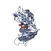
- Assembly
| Deposited unit | 
| ||||||||
|---|---|---|---|---|---|---|---|---|---|
| 1 |
| ||||||||
| Unit cell |
|
-Components
| #1: Antibody | Mass: 14350.981 Da / Num. of mol.: 2 Source method: isolated from a genetically manipulated source Source: (gene. exp.)  #2: Chemical | ChemComp-SO4 / |   Mass: 96.063 Da / Num. of mol.: 1 / Source method: obtained synthetically / Formula: SO4 Mass: 96.063 Da / Num. of mol.: 1 / Source method: obtained synthetically / Formula: SO4#3: Water | ChemComp-HOH / |  Mass: 18.015 Da / Num. of mol.: 103 / Source method: isolated from a natural source / Formula: H2O Mass: 18.015 Da / Num. of mol.: 103 / Source method: isolated from a natural source / Formula: H2OCompound details | SEQUENCE REFLECTS C22A, C99V MUTATIONS AGAINST WT PROTEIN. NO UNP MATCH IS FOUND, THEREFORE THE ...SEQUENCE REFLECTS C22A, C99V MUTATIONS AGAINST WT PROTEIN. NO UNP MATCH IS FOUND, THEREFORE THE SEQUENCE IS SELF REFERENCED | |
|---|
-Experimental details
-Experiment
| Experiment | Method: X-RAY DIFFRACTION |
|---|
- Sample preparation
| Crystal | Density Matthews: 2.43 Å3/Da / Density % sol: 49.47 % / Description: rod-shaped |
|---|---|
| Crystal grow | Temperature: 290 K / Method: vapor diffusion, hanging drop / pH: 8 Details: 0.1 M Tris pH 8.0, 1.6 M Ammonium Sulfate [Hampton Research Ammonium Sulfate Grid Screen, condition B5] |
-Data collection
| Diffraction | Mean temperature: 150 K |
|---|---|
| Diffraction source | Source: ROTATING ANODE / Type: BRUKER AXS MICROSTAR-H / Wavelength: 1.54 Å |
| Detector | Type: BRUKER SMART 6000 / Detector: CCD / Date: Sep 4, 2013 |
| Radiation | Protocol: SINGLE WAVELENGTH / Monochromatic (M) / Laue (L): M / Scattering type: x-ray |
| Radiation wavelength | Wavelength: 1.54 Å / Relative weight: 1 |
| Reflection | Resolution: 2→58.05 Å / Num. obs: 18197 / % possible obs: 94.3 % / Redundancy: 10.57 % / Biso Wilson estimate: 15.3 Å2 / Rmerge(I) obs: 0.1038 / Rsym value: 0.054 / Net I/σ(I): 16.86 |
| Reflection shell | Resolution: 2→2.09 Å / Redundancy: 2.29 % / Rmerge(I) obs: 0.3556 / Mean I/σ(I) obs: 3.32 / % possible all: 71.8 |
- Processing
| Software | Name: REFMAC / Version: 5.6.0117 / Classification: refinement | ||||||||||||||||||||||||||||||||||||||||||||||||||||||||||||||||||||||||||||||||||||||||||||||||||||||||||||||||||||||||||||||||||||||||||||||||||||||||||||||||||||||||||||||||||||||
|---|---|---|---|---|---|---|---|---|---|---|---|---|---|---|---|---|---|---|---|---|---|---|---|---|---|---|---|---|---|---|---|---|---|---|---|---|---|---|---|---|---|---|---|---|---|---|---|---|---|---|---|---|---|---|---|---|---|---|---|---|---|---|---|---|---|---|---|---|---|---|---|---|---|---|---|---|---|---|---|---|---|---|---|---|---|---|---|---|---|---|---|---|---|---|---|---|---|---|---|---|---|---|---|---|---|---|---|---|---|---|---|---|---|---|---|---|---|---|---|---|---|---|---|---|---|---|---|---|---|---|---|---|---|---|---|---|---|---|---|---|---|---|---|---|---|---|---|---|---|---|---|---|---|---|---|---|---|---|---|---|---|---|---|---|---|---|---|---|---|---|---|---|---|---|---|---|---|---|---|---|---|---|---|
| Refinement | Method to determine structure: MOLECULAR REPLACEMENT Starting model: 4TYU Resolution: 2→58.05 Å / Cor.coef. Fo:Fc: 0.93 / Cor.coef. Fo:Fc free: 0.892 / Cross valid method: THROUGHOUT / ESU R: 0.201 / ESU R Free: 0.175 / Stereochemistry target values: MAXIMUM LIKELIHOOD / Details: HYDROGENS HAVE BEEN USED IF PRESENT IN THE INPUT
| ||||||||||||||||||||||||||||||||||||||||||||||||||||||||||||||||||||||||||||||||||||||||||||||||||||||||||||||||||||||||||||||||||||||||||||||||||||||||||||||||||||||||||||||||||||||
| Solvent computation | Ion probe radii: 0.8 Å / Shrinkage radii: 0.8 Å / VDW probe radii: 1.2 Å / Solvent model: MASK | ||||||||||||||||||||||||||||||||||||||||||||||||||||||||||||||||||||||||||||||||||||||||||||||||||||||||||||||||||||||||||||||||||||||||||||||||||||||||||||||||||||||||||||||||||||||
| Displacement parameters | Biso mean: 14.252 Å2
| ||||||||||||||||||||||||||||||||||||||||||||||||||||||||||||||||||||||||||||||||||||||||||||||||||||||||||||||||||||||||||||||||||||||||||||||||||||||||||||||||||||||||||||||||||||||
| Refinement step | Cycle: 1 / Resolution: 2→58.05 Å
| ||||||||||||||||||||||||||||||||||||||||||||||||||||||||||||||||||||||||||||||||||||||||||||||||||||||||||||||||||||||||||||||||||||||||||||||||||||||||||||||||||||||||||||||||||||||
| Refine LS restraints |
|