 Movie
Movie Controller

[English] 日本語
 Yorodumi
- PDB-2hr6: Crystal structure of dUTPase in complex with substrate analogue d... -
+ Open data

- Basic information
| Entry | Database: PDB / ID: 2hr6 | ||||||
|---|---|---|---|---|---|---|---|
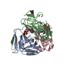
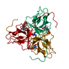
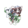
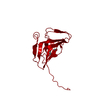
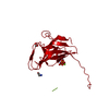
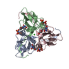
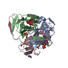
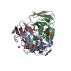
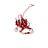
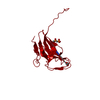
| Title | Crystal structure of dUTPase in complex with substrate analogue dUDP and manganese | ||||||
 Components Components | Deoxyuridine 5'-triphosphate nucleotidohydrolase | ||||||
 Keywords Keywords | HYDROLASE / JELLY ROLL / ENZYME-LIGAND-metal COMPLEX | ||||||
| Function / homology |  Function and homology information Function and homology informationdUTP catabolic process / dUMP biosynthetic process / dUTP diphosphatase / dUTP diphosphatase activity / protein homotrimerization / magnesium ion binding / protein-containing complex / identical protein binding / cytosol Similarity search - Function | ||||||
| Biological species |  | ||||||
| Method |  X-RAY DIFFRACTION / SYNCHROTRON / MOLECULAR REPLACEMENT / Resolution: 1.84 Å X-RAY DIFFRACTION / SYNCHROTRON / MOLECULAR REPLACEMENT / Resolution: 1.84 Å | ||||||
 Authors Authors | Barabas, O. / Kovari, J. / Tapai, R. / Vertessy, B.G. | ||||||
 Citation Citation | Journal: Proteins / Year: 2008 Title: Methylene substitution at the alpha-beta bridging position within the phosphate chain of dUDP profoundly perturbs ligand accommodation into the dUTPase active site. Authors: Kovari, J. / Barabas, O. / Varga, B. / Bekesi, A. / Tolgyesi, F. / Fidy, J. / Nagy, J. / Vertessy, B.G. #1: Journal: J.Biol.Chem. / Year: 2004Title: Structural Insights into the Catalytic Mechanism of Phosphate Ester Hydrolysis by dUTPase. Authors: Barabas, O. / Pongracz, V. / Kovari, J. / Wilmanns, M. / Vertessy, B.G. #2: Journal: Acta Crystallogr.,Sect.D / Year: 2001Title: Atomic Resolution Structure of Escherichia Coli Dutpase Determined Ab Initio Authors: Gonzalez, A. / Larsson, G. / Persson, R. / Cedergren-Zeppezauer, E. | ||||||
| History |
|
- Structure visualization
| Structure viewer | Molecule: MolmilJmol/JSmol |
|---|
- Downloads & links
-Download
| PDBx/mmCIF format | 2hr6.cif.gz | 47.5 KB | Display | PDBx/mmCIF format |
|---|---|---|---|---|
| PDB format | pdb2hr6.ent.gz | 31.7 KB | Display | PDB format |
| PDBx/mmJSON format | 2hr6.json.gz | Tree view | PDBx/mmJSON format | |
| Others |  Other downloads Other downloads |
-Validation report
| Arichive directory | https://data.pdbj.org/pub/pdb/validation_reports/hr/2hr6ftp://data.pdbj.org/pub/pdb/validation_reports/hr/2hr6 | HTTPS FTP |
|---|
-Related structure data
| Related structure data |  2hrmC 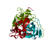 1euwS S: Starting model for refinement C: citing same article ( |
|---|---|
| Similar structure data |
-Links
 PDBj
PDBj
- Assembly
| Deposited unit | 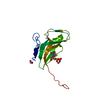
| ||||||||
|---|---|---|---|---|---|---|---|---|---|
| 1 | 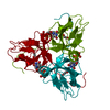
| ||||||||
| Unit cell |
| ||||||||
| Components on special symmetry positions |
| ||||||||
| Details | The biological assembly is a trimer generated from the monomer in the asymmetric unit by the operations: 1-y,1+x-y,z and -x+y,1-x,z |
-Components
| #1: Protein | Mass: 16302.615 Da / Num. of mol.: 1 Source method: isolated from a genetically manipulated source Source: (gene. exp.) |
|---|---|
| #2: Chemical | ChemComp-MN /   Mass: 54.938 Da / Num. of mol.: 1 / Source method: obtained synthetically / Formula: Mn Mass: 54.938 Da / Num. of mol.: 1 / Source method: obtained synthetically / Formula: Mn |
| #3: Chemical | ChemComp-DUD /   Mass: 388.162 Da / Num. of mol.: 1 / Source method: obtained synthetically / Formula: C9H14N2O11P2 Mass: 388.162 Da / Num. of mol.: 1 / Source method: obtained synthetically / Formula: C9H14N2O11P2 |
| #4: Chemical | ChemComp-EDO /   Mass: 62.068 Da / Num. of mol.: 1 / Source method: obtained synthetically / Formula: C2H6O2 Mass: 62.068 Da / Num. of mol.: 1 / Source method: obtained synthetically / Formula: C2H6O2 |
| #5: Water | ChemComp-HOH /  Mass: 18.015 Da / Num. of mol.: 156 / Source method: isolated from a natural source / Formula: H2O Mass: 18.015 Da / Num. of mol.: 156 / Source method: isolated from a natural source / Formula: H2O |
-Experimental details
-Experiment
| Experiment | Method: X-RAY DIFFRACTION / Number of used crystals: 1 |
|---|
- Sample preparation
| Crystal | Density Matthews: 2.49 Å3/Da / Density % sol: 50.61 % |
|---|---|
| Crystal grow | Temperature: 293 K / Method: vapor diffusion, hanging drop / pH: 7.8 Details: 20% PEG 3350, 0.2M SODIUM ACETATE, 0.1M TRIS, pH 7.8, VAPOR DIFFUSION, HANGING DROP, temperature 293K |
-Data collection
| Diffraction | Mean temperature: 100 K |
|---|---|
| Diffraction source | Source: SYNCHROTRON / Site: EMBL/DESY, HAMBURG  / Beamline: X11 / Wavelength: 0.811 Å / Beamline: X11 / Wavelength: 0.811 Å |
| Detector | Type: MAR CCD 165 mm / Detector: CCD / Date: Mar 17, 2002 / Details: mirror |
| Radiation | Monochromator: TRIANGULAR MONOCHROMATOR / Protocol: SINGLE WAVELENGTH / Monochromatic (M) / Laue (L): M / Scattering type: x-ray |
| Radiation wavelength | Wavelength: 0.811 Å / Relative weight: 1 |
| Reflection | Resolution: 1.84→24.9 Å / Num. all: 14703 / Num. obs: 14703 / % possible obs: 98 % / Observed criterion σ(I): -3 / Redundancy: 12.6 % / Biso Wilson estimate: 22.35 Å2 / Rsym value: 0.064 / Net I/σ(I): 10.8 |
| Reflection shell | Resolution: 1.84→1.89 Å / Redundancy: 10.1 % / Mean I/σ(I) obs: 1.3 / Num. unique all: 815 / Rsym value: 0.558 / % possible all: 98 |
- Processing
| Software |
| |||||||||||||||||||||||||||||||||||||||||||||||||||||||||||||||||||||||||||||||||||||||||||||||||||||||||||||||||||||||||||||
|---|---|---|---|---|---|---|---|---|---|---|---|---|---|---|---|---|---|---|---|---|---|---|---|---|---|---|---|---|---|---|---|---|---|---|---|---|---|---|---|---|---|---|---|---|---|---|---|---|---|---|---|---|---|---|---|---|---|---|---|---|---|---|---|---|---|---|---|---|---|---|---|---|---|---|---|---|---|---|---|---|---|---|---|---|---|---|---|---|---|---|---|---|---|---|---|---|---|---|---|---|---|---|---|---|---|---|---|---|---|---|---|---|---|---|---|---|---|---|---|---|---|---|---|---|---|---|
| Refinement | Method to determine structure: MOLECULAR REPLACEMENT Starting model: 1EUW Resolution: 1.84→24.9 Å / Cor.coef. Fo:Fc: 0.968 / Cor.coef. Fo:Fc free: 0.963 / SU B: 4.48 / SU ML: 0.064 / TLS residual ADP flag: LIKELY RESIDUAL / Cross valid method: THROUGHOUT / ESU R: 0.103 / ESU R Free: 0.096 / Stereochemistry target values: MAXIMUM LIKELIHOOD / Details: HYDROGENS HAVE BEEN ADDED IN THE RIDING POSITIONS
| |||||||||||||||||||||||||||||||||||||||||||||||||||||||||||||||||||||||||||||||||||||||||||||||||||||||||||||||||||||||||||||
| Solvent computation | Ion probe radii: 0.8 Å / Shrinkage radii: 0.8 Å / VDW probe radii: 1.2 Å / Solvent model: BABINET MODEL WITH MASK | |||||||||||||||||||||||||||||||||||||||||||||||||||||||||||||||||||||||||||||||||||||||||||||||||||||||||||||||||||||||||||||
| Displacement parameters | Biso mean: 22.763 Å2
| |||||||||||||||||||||||||||||||||||||||||||||||||||||||||||||||||||||||||||||||||||||||||||||||||||||||||||||||||||||||||||||
| Refinement step | Cycle: LAST / Resolution: 1.84→24.9 Å
| |||||||||||||||||||||||||||||||||||||||||||||||||||||||||||||||||||||||||||||||||||||||||||||||||||||||||||||||||||||||||||||
| Refine LS restraints |
| |||||||||||||||||||||||||||||||||||||||||||||||||||||||||||||||||||||||||||||||||||||||||||||||||||||||||||||||||||||||||||||
| LS refinement shell | Resolution: 1.842→1.89 Å / Total num. of bins used: 20
| |||||||||||||||||||||||||||||||||||||||||||||||||||||||||||||||||||||||||||||||||||||||||||||||||||||||||||||||||||||||||||||
| Refinement TLS params. | Method: refined / Origin x: 0.1866 Å / Origin y: 31.3803 Å / Origin z: 4.8255 Å
| |||||||||||||||||||||||||||||||||||||||||||||||||||||||||||||||||||||||||||||||||||||||||||||||||||||||||||||||||||||||||||||
| Refinement TLS group |
|
