 Movie
Movie Controller

[English] 日本語
 Yorodumi
- PDB-2hlf: Structure of the Escherichis coli ClC chloride channel Y445E muta... -
+ Open data

- Basic information
| Entry | Database: PDB / ID: 2hlf | ||||||
|---|---|---|---|---|---|---|---|
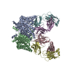
| Title | Structure of the Escherichis coli ClC chloride channel Y445E mutant and Fab complex | ||||||
 Components Components |
| ||||||
 Keywords Keywords | PROTON TRANSPORT / MEMBRANE PROTEIN / ClC family of channels and transpoters / H+/Cl- antiporter / Fab complex | ||||||
| Function / homology |  Function and homology information Function and homology informationcellular stress response to acidic pH / chloride:proton antiporter activity / voltage-gated chloride channel activity / proton transmembrane transport / chloride transmembrane transport / identical protein binding / plasma membrane Similarity search - Function | ||||||
| Biological species |   | ||||||
| Method |  X-RAY DIFFRACTION / SYNCHROTRON / MOLECULAR REPLACEMENT / Resolution: 3.3 Å X-RAY DIFFRACTION / SYNCHROTRON / MOLECULAR REPLACEMENT / Resolution: 3.3 Å | ||||||
 Authors Authors | Accardi, A. / Lobet, S. / Williams, C. / Miller, C. / Dutzler, R. | ||||||
 Citation Citation | Journal: J.Mol.Biol. / Year: 2006 Title: Synergism Between Halide Binding and Proton Transport in a CLC-type Exchanger Authors: Accardi, A. / Lobet, S. / Williams, C. / Miller, C. / Dutzler, R. | ||||||
| History |
|
- Structure visualization
| Structure viewer | Molecule: MolmilJmol/JSmol |
|---|
- Downloads & links
-Download
| PDBx/mmCIF format | 2hlf.cif.gz | 336.5 KB | Display | PDBx/mmCIF format |
|---|---|---|---|---|
| PDB format | pdb2hlf.ent.gz | 272 KB | Display | PDB format |
| PDBx/mmJSON format | 2hlf.json.gz | Tree view | PDBx/mmJSON format | |
| Others |  Other downloads Other downloads |
-Validation report
| Summary document | 2hlf_validation.pdf.gz | 485.6 KB | Display | wwPDB validaton report |
|---|---|---|---|---|
| Full document | 2hlf_full_validation.pdf.gz | 573.8 KB | Display | |
| Data in XML | 2hlf_validation.xml.gz | 70.4 KB | Display | |
| Data in CIF | 2hlf_validation.cif.gz | 94.8 KB | Display | |
| Arichive directory | https://data.pdbj.org/pub/pdb/validation_reports/hl/2hlfftp://data.pdbj.org/pub/pdb/validation_reports/hl/2hlf | HTTPS FTP |
-Related structure data
| Related structure data |  2ht2C  2ht3C 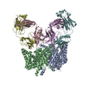 2ht4C  2htkC 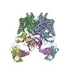 2htlC 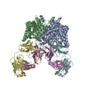 1otsS S: Starting model for refinement C: citing same article ( |
|---|---|
| Similar structure data |


-Links
 PDBj
PDBj


- Assembly
| Deposited unit | 
| ||||||||||||||||||
|---|---|---|---|---|---|---|---|---|---|---|---|---|---|---|---|---|---|---|---|
| 1 |
| ||||||||||||||||||
| Unit cell |
| ||||||||||||||||||
| Noncrystallographic symmetry (NCS) | NCS domain:
NCS domain segments: Component-ID: 1 / Ens-ID: 1 / Beg auth comp-ID: ARG / Beg label comp-ID: ARG / End auth comp-ID: ALA / End label comp-ID: ALA / Refine code: 2 / Auth seq-ID: 18 - 458 / Label seq-ID: 2 - 442
|
-Components
| #1: Protein | Mass: 47323.969 Da / Num. of mol.: 2 / Mutation: Y445E Source method: isolated from a genetically manipulated source Details: Each subunit of the mutant has a single Br- ion bound in the selectivity filter Source: (gene. exp.) #2: Antibody | Mass: 23693.918 Da / Num. of mol.: 2 / Source method: isolated from a natural source / Source: (natural) #3: Antibody | Mass: 23088.443 Da / Num. of mol.: 2 / Source method: isolated from a natural source / Source: (natural) #4: Chemical |   Mass: 79.904 Da / Num. of mol.: 2 / Source method: obtained synthetically / Formula: Br Mass: 79.904 Da / Num. of mol.: 2 / Source method: obtained synthetically / Formula: BrHas protein modification | Y | |
|---|
-Experimental details
-Experiment
| Experiment | Method: X-RAY DIFFRACTION / Number of used crystals: 1 |
|---|
- Sample preparation
| Crystal | Density Matthews: 3.87 Å3/Da / Density % sol: 68.26 % |
|---|---|
| Crystal grow | Temperature: 293 K / Method: vapor diffusion, sitting drop / pH: 9.5 Details: 34% peg 200/300 1:2, 50mM glycin, 150mM NaBr, pH 9.5, VAPOR DIFFUSION, SITTING DROP, temperature 293K |
-Data collection
| Diffraction | Mean temperature: 100 K |
|---|---|
| Diffraction source | Source: SYNCHROTRON / Site: SLS  / Beamline: X06SA / Wavelength: 0.91921 Å / Beamline: X06SA / Wavelength: 0.91921 Å |
| Detector | Type: MARRESEARCH / Detector: CCD / Date: Jun 24, 2005 |
| Radiation | Monochromator: LN2 cooled fixed-exit Si(111) monochromator / Protocol: SINGLE WAVELENGTH / Monochromatic (M) / Laue (L): M / Scattering type: x-ray |
| Radiation wavelength | Wavelength: 0.91921 Å / Relative weight: 1 |
| Reflection | Resolution: 3.3→50 Å / Num. all: 44455 / Num. obs: 42596 / % possible obs: 95.8 % / Observed criterion σ(I): 1 / Biso Wilson estimate: 109 Å2 |
| Reflection shell | Resolution: 3.3→3.42 Å / % possible all: 81.7 |
- Processing
| Software |
| |||||||||||||||||||||||||
|---|---|---|---|---|---|---|---|---|---|---|---|---|---|---|---|---|---|---|---|---|---|---|---|---|---|---|
| Refinement | Method to determine structure: MOLECULAR REPLACEMENT Starting model: 1OTS Resolution: 3.3→40 Å / Cor.coef. Fo:Fc: 0.921 / Cor.coef. Fo:Fc free: 0.902 / Isotropic thermal model: restrained / Cross valid method: THROUGHOUT / σ(F): 0 / ESU R: 0.543 / ESU R Free: 0.597 / Stereochemistry target values: Engh & Huber
| |||||||||||||||||||||||||
| Solvent computation | Ion probe radii: 0.8 Å / Shrinkage radii: 0.8 Å / VDW probe radii: 1.2 Å / Solvent model: MASK | |||||||||||||||||||||||||
| Displacement parameters | Biso mean: 128.436 Å2
| |||||||||||||||||||||||||
| Refinement step | Cycle: LAST / Resolution: 3.3→40 Å
| |||||||||||||||||||||||||
| Refine LS restraints |
| |||||||||||||||||||||||||
| LS refinement shell | Resolution: 3.3→3.385 Å / Total num. of bins used: 20
|
