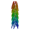
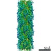
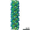
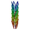
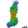
Journal: J Mol Biol / Year: 2006 Title: The structure of a filamentous bacteriophage. Authors: Ying A Wang / Xiong Yu / Stacy Overman / Masamichi Tsuboi / George J Thomas / Edward H Egelman / Abstract: Many thin helical polymers, including bacterial pili and filamentous bacteriophage, have been seen as refractory to high-resolution studies by electron microscopy. Studies of the quaternary structure ...Many thin helical polymers, including bacterial pili and filamentous bacteriophage, have been seen as refractory to high-resolution studies by electron microscopy. Studies of the quaternary structure of such filaments have depended upon techniques such as modeling or X-ray fiber diffraction, given that direct visualization of the subunit organization has not been possible. We report the first image reconstruction of a filamentous virus, bacteriophage fd, by cryoelectron microscopy. Although these thin ( approximately 70 A in diameter) rather featureless filaments scatter weakly, we have been able to achieve a nominal resolution of approximately 8 A using an iterative helical reconstruction procedure. We show that two different conformations of the virus exist, and that in both states the subunits are packed differently than in conflicting models previously proposed on the basis of X-ray fiber diffraction or solid-state NMR studies. A significant fraction of the population of wild-type fd is either disordered or in multiple conformational states, while in the presence of the Y21M mutation, this heterogeneity is greatly reduced, consistent with previous observations. These results show that new computational approaches to helical reconstruction can greatly extend the ability to visualize heterogeneous protein polymers at a reasonably high resolution.
Helical symmetry: (Circular symmetry: 5 / Dyad axis: no / N subunits divisor: 1 / Num. of operations: 55 / Rise per n subunits: 17.4 Å / Rotation per n subunits: -34.616 °)
Details
helical polymer
-
Components
#1: Protein/peptide
CoatproteinB / Major coat protein
Mass: 5244.000 Da / Num. of mol.: 1 Source method: isolated from a genetically manipulated source Source: (gene. exp.) Enterobacteria phage fd (virus) / Genus: Inovirus / Species: Enterobacteria phage M13 / Production host: Escherichia coli (E. coli) / References: UniProt: P69539
-
Experimental details
-
Experiment
Experiment
Method: ELECTRON MICROSCOPY
EM experiment
Aggregation state: FILAMENT / 3D reconstruction method: helical reconstruction
-
Sample preparation
Component
ID
Name
Type
Parent-ID
Details
1
Bacteriophagefd
VIRUS
0
2
CoatproteinB
1
viralcapsid
Specimen
Embedding applied: NO / Shadowing applied: NO / Staining applied: NO / Vitrification applied: YES
Vitrification
Cryogen name: ETHANE / Method: Blot for 2 seconds before plunging
-
Electron microscopy imaging
Experimental equipment
Model: Tecnai F20 / Image courtesy: FEI Company
Microscopy
Model: FEI TECNAI F20
Electron gun
Electron source: FIELD EMISSION GUN / Accelerating voltage: 200 kV / Illumination mode: FLOOD BEAM
Electron lens
Mode: BRIGHT FIELD / Nominal magnification: 50000 X / Nominal defocus max: 3800 nm / Nominal defocus min: 1400 nm
Specimen holder
Specimen holder type: Gatan
Image recording
Film or detector model: KODAK SO-163 FILM
Radiation
Protocol: SINGLE WAVELENGTH / Monochromatic (M) / Laue (L): M
Radiation wavelength
Relative weight: 1
-
Processing
EM software
Name: IHRSR / Category: 3D reconstruction
CTF correction
Details: phase flipping of entire images
3D reconstruction
Method: IHRSR / Resolution: 8 Å / Resolution method: FSC 0.5 CUT-OFF / Num. of particles: 16367 / Nominal pixel size: 2.4 Å / Actual pixel size: 2.4 Å / Magnification calibration: TMV Details: The C-N bond distance is 0.09 A between GLN15 and ALA16, 0.12 A between ALA35 and THR36 and 0.44 A between ALA25 and TRP26 Symmetry type: HELICAL
Refinement step
Cycle: LAST
Protein
Nucleic acid
Ligand
Solvent
Total
Num. atoms
315
0
0
0
315
+
About Yorodumi
-
News
-
Feb 9, 2022. New format data for meta-information of EMDB entries
New format data for meta-information of EMDB entries
Version 3 of the EMDB header file is now the official format.
The previous official version 1.9 will be removed from the archive.
In the structure databanks used in Yorodumi, some data are registered as the other names, "COVID-19 virus" and "2019-nCoV". Here are the details of the virus and the list of structure data.
Jan 31, 2019. EMDB accession codes are about to change! (news from PDBe EMDB page)
EMDB accession codes are about to change! (news from PDBe EMDB page)
The allocation of 4 digits for EMDB accession codes will soon come to an end. Whilst these codes will remain in use, new EMDB accession codes will include an additional digit and will expand incrementally as the available range of codes is exhausted. The current 4-digit format prefixed with “EMD-” (i.e. EMD-XXXX) will advance to a 5-digit format (i.e. EMD-XXXXX), and so on. It is currently estimated that the 4-digit codes will be depleted around Spring 2019, at which point the 5-digit format will come into force.
The EM Navigator/Yorodumi systems omit the EMD- prefix.
Related info.:Q: What is EMD? / ID/Accession-code notation in Yorodumi/EM Navigator
Yorodumi is a browser for structure data from EMDB, PDB, SASBDB, etc.
This page is also the successor to EM Navigator detail page, and also detail information page/front-end page for Omokage search.
The word "yorodu" (or yorozu) is an old Japanese word meaning "ten thousand". "mi" (miru) is to see.
Related info.:EMDB / PDB / SASBDB / Comparison of 3 databanks / Yorodumi Search / Aug 31, 2016. New EM Navigator & Yorodumi / Yorodumi Papers / Jmol/JSmol / Function and homology information / Changes in new EM Navigator and Yorodumi
 Movie
Movie Controller


 Open data
 Basic information
 Components
 Keywords
 Function and homology information
 Enterobacteria phage fd (virus)
 Authors
 Citation
 Structure visualization
 Downloads & links
 Other downloads
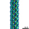

 PDBj
PDBj Assembly


 Sample preparation
 Electron microscopy imaging
 FIELD EMISSION GUN / Accelerating voltage: 200 kV / Illumination mode: FLOOD BEAM
 Processing