- PDB-2hi0: Crystal structure of putative phosphoglycolate phosphatase (YP_61... -
+
Open data
ID or keywords:
Loading...
-
Basic information
Entry
Database: PDB / ID: 2hi0
Title
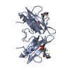
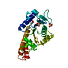
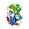
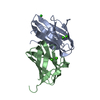
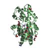
Crystal structure of putative phosphoglycolate phosphatase (YP_619066.1) from Lactobacillus delbrueckii subsp. bulgaricus ATCC BAA-365 at 1.51 A resolution
Components
Putative phosphoglycolate phosphatase
Keywords
HYDROLASE / YP_619066.1 / putative / Structural Genomics / Joint Center for Structural Genomics / JCSG / Protein Structure Initiative / PSI
Function / homology
Function and homology information
phosphoglycolate phosphatase / phosphoglycolate phosphatase activity / DNA repair / cytosol Similarity search - Function
: / Haloacid dehalogenase-like hydrolase / Haloacid dehalogenase-like hydrolase / Putative phosphatase; domain 2 / Phosphoglycolate phosphatase-like, domain 2 / HAD hydrolase, subfamily IA / HAD superfamily/HAD-like / HAD superfamily / HAD-like superfamily / DNA polymerase; domain 1 ...: / Haloacid dehalogenase-like hydrolase / Haloacid dehalogenase-like hydrolase / Putative phosphatase; domain 2 / Phosphoglycolate phosphatase-like, domain 2 / HAD hydrolase, subfamily IA / HAD superfamily/HAD-like / HAD superfamily / HAD-like superfamily / DNA polymerase; domain 1 / Rossmann fold / Orthogonal Bundle / 3-Layer(aba) Sandwich / Mainly Alpha / Alpha Beta Similarity search - Domain/homology
SEQUENCE THE SEQUENCE DIFFERENCE ARISES BECAUSE THE PROTEIN WAS OBTAINED FROM A DIFFERENT ...SEQUENCE THE SEQUENCE DIFFERENCE ARISES BECAUSE THE PROTEIN WAS OBTAINED FROM A DIFFERENT SUBSPECIES (ATCC BAA-365). THE CONSTRUCT WAS EXPRESSED WITH A PURIFICATION TAG MGSDKIHHHHHHENLYFQG. THE TAG WAS REMOVED WITH TEV PROTEASE LEAVING ONLY A GLYCINE (0) FOLLOWED BY THE TARGET SEQUENCE.
Resolution: 1.51→47.727 Å / Num. obs: 72271 / % possible obs: 95.8 % / Redundancy: 3.61 % / Biso Wilson estimate: 19.143 Å2 / Rmerge(I) obs: 0.101 / Net I/σ(I): 8.75
Reflection shell
Diffraction-ID: 1
Resolution (Å)
Highest resolution (Å)
Redundancy (%)
Rmerge(I) obs
Mean I/σ(I) obs
Num. measured obs
Num. unique all
% possible all
1.51-1.56
2.77
0.537
2.1
15640
5575
82.6
1.56-1.63
0.515
2.6
27140
7550
93.2
1.63-1.7
0.448
3.1
23957
6439
94.6
1.7-1.79
0.365
3.8
25809
6958
95.7
1.79-1.9
0.273
4.9
25745
6904
96.6
1.9-2.05
0.184
7.1
26987
7235
97.6
2.05-2.25
0.121
10
26070
7015
98.9
2.25-2.58
0.092
12.6
27493
7365
99.1
2.58
0.068
15.7
27229
7316
99.3
-
Phasing
Phasing
Method: MAD
-
Processing
Software
Name
Version
Classification
NB
MolProbity
3beta29
modelbuilding
REFMAC
5.2.0019
refinement
XSCALE
datascaling
PDB_EXTRACT
2
dataextraction
XDS
datareduction
SHELXD
phasing
autoSHARP
phasing
Refinement
Method to determine structure: MAD / Resolution: 1.51→47.727 Å / Cor.coef. Fo:Fc: 0.969 / Cor.coef. Fo:Fc free: 0.953 / SU B: 2.969 / SU ML: 0.055 / TLS residual ADP flag: LIKELY RESIDUAL / Cross valid method: THROUGHOUT / σ(F): 0 / ESU R: 0.072 / ESU R Free: 0.075 Stereochemistry target values: MAXIMUM LIKELIHOOD WITH PHASES Details: (1) HYDROGENS HAVE BEEN ADDED IN THE RIDING POSITIONS. (2) A MET-INHIBITION PROTOCOL WAS USED FOR SELENOMETHIONINE INCORPORATION DURING PROTEIN EXPRESSION. THE OCCUPANCY OF THE SE ATOMS IN ...Details: (1) HYDROGENS HAVE BEEN ADDED IN THE RIDING POSITIONS. (2) A MET-INHIBITION PROTOCOL WAS USED FOR SELENOMETHIONINE INCORPORATION DURING PROTEIN EXPRESSION. THE OCCUPANCY OF THE SE ATOMS IN THE MSE RESIDUES WAS REDUCED TO 0.75 TO ACCOUNT FOR THE REDUCED SCATTERING POWER DUE TO PARTIAL S-MET INCORPORATION. (3) NA, CL, ACT AND EDO WERE MODELLED BASED ON CRYSTALLIZATION CONDITIONS AND THEIR GEOMETRY. (4) ATOM RECORDS CONTAIN RESIDUAL B FACTORS ONLY.
Rfactor
Num. reflection
% reflection
Selection details
Rfree
0.19
3644
5 %
RANDOM
Rwork
0.157
-
-
-
all
0.158
-
-
-
obs
0.158
72257
99.17 %
-
Solvent computation
Ion probe radii: 0.8 Å / Shrinkage radii: 0.8 Å / VDW probe radii: 1.2 Å / Solvent model: MASK
Displacement parameters
Biso mean: 11.423 Å2
Baniso -1
Baniso -2
Baniso -3
1-
-0.07 Å2
0 Å2
0.19 Å2
2-
-
0.17 Å2
0 Å2
3-
-
-
-0.05 Å2
Refinement step
Cycle: LAST / Resolution: 1.51→47.727 Å
Protein
Nucleic acid
Ligand
Solvent
Total
Num. atoms
3675
0
81
637
4393
Refine LS restraints
Refine-ID
Type
Dev ideal
Dev ideal target
Number
X-RAY DIFFRACTION
r_bond_refined_d
0.017
0.022
3881
X-RAY DIFFRACTION
r_bond_other_d
0.002
0.02
2642
X-RAY DIFFRACTION
r_angle_refined_deg
1.592
1.97
5239
X-RAY DIFFRACTION
r_angle_other_deg
0.99
3
6458
X-RAY DIFFRACTION
r_dihedral_angle_1_deg
5.487
5
499
X-RAY DIFFRACTION
r_dihedral_angle_2_deg
34.547
24.588
170
X-RAY DIFFRACTION
r_dihedral_angle_3_deg
13.063
15
653
X-RAY DIFFRACTION
r_dihedral_angle_4_deg
15.444
15
22
X-RAY DIFFRACTION
r_chiral_restr
0.093
0.2
592
X-RAY DIFFRACTION
r_gen_planes_refined
0.007
0.02
4355
X-RAY DIFFRACTION
r_gen_planes_other
0.001
0.02
765
X-RAY DIFFRACTION
r_nbd_refined
0.229
0.2
791
X-RAY DIFFRACTION
r_nbd_other
0.196
0.2
2810
X-RAY DIFFRACTION
r_nbtor_refined
0.182
0.2
1920
X-RAY DIFFRACTION
r_nbtor_other
0.086
0.2
1893
X-RAY DIFFRACTION
r_xyhbond_nbd_refined
0.151
0.2
440
X-RAY DIFFRACTION
r_metal_ion_refined
0.208
0.2
7
X-RAY DIFFRACTION
r_symmetry_vdw_refined
0.176
0.2
24
X-RAY DIFFRACTION
r_symmetry_vdw_other
0.289
0.2
64
X-RAY DIFFRACTION
r_symmetry_hbond_refined
0.177
0.2
54
X-RAY DIFFRACTION
r_mcbond_it
1.707
3
2443
X-RAY DIFFRACTION
r_mcbond_other
0.532
3
987
X-RAY DIFFRACTION
r_mcangle_it
2.632
5
3944
X-RAY DIFFRACTION
r_scbond_it
4.43
8
1458
X-RAY DIFFRACTION
r_scangle_it
6.563
11
1289
LS refinement shell
Resolution: 1.51→1.549 Å / Total num. of bins used: 20
Rfactor
Num. reflection
% reflection
Rfree
0.32
245
-
Rwork
0.25
4637
-
obs
-
4882
90.69 %
Refinement TLS params.
Method: refined / Refine-ID: X-RAY DIFFRACTION
ID
L11 (°2)
L12 (°2)
L13 (°2)
L22 (°2)
L23 (°2)
L33 (°2)
S11 (Å °)
S12 (Å °)
S13 (Å °)
S21 (Å °)
S22 (Å °)
S23 (Å °)
S31 (Å °)
S32 (Å °)
S33 (Å °)
T11 (Å2)
T12 (Å2)
T13 (Å2)
T22 (Å2)
T23 (Å2)
T33 (Å2)
Origin x (Å)
Origin y (Å)
Origin z (Å)
1
0.7617
-0.3193
-0.3609
0.782
-0.1828
0.8631
-0.0221
-0.0479
-0.0486
0.0281
-0.0021
-0.0233
0.0765
0.1146
0.0242
-0.0013
0.0226
0.0085
-0.0133
0.0025
-0.011
6.2323
6.8739
36.148
2
0.8183
-0.1945
-0.0053
0.694
0.2826
1.2497
-0.024
0.029
-0.0494
0.0125
0.0182
0.0747
0.0058
-0.0664
0.0058
-0.045
-0.0022
0.0045
-0.0401
0.0043
-0.0269
-14.1639
17.1264
37.0173
3
0.6629
-0.1499
-0.1235
0.3474
0.0993
1.0725
0.0047
0.0287
-0.0163
-0.0185
0.0152
0.0035
-0.0255
-0.0342
-0.02
-0.0332
-0.0057
-0.0032
-0.0238
-0.0043
-0.0155
10.9925
-14.4616
9.2787
4
0.42
-0.0293
-0.2759
0.4741
-0.1519
1.294
-0.0176
-0.0103
-0.0392
0.0117
-0.0127
-0.0457
-0.0243
0.0386
0.0303
-0.0505
-0.0053
-0.007
-0.026
-0.0008
-0.0223
30.5584
-3.4708
15.4184
Refinement TLS group
Refine-ID: X-RAY DIFFRACTION / Selection: ALL
ID
Refine TLS-ID
Auth asym-ID
Label asym-ID
Auth seq-ID
Label seq-ID
1
1
A
A
0 - 16
1 - 17
2
1
A
A
109 - 239
110 - 240
3
2
A
A
17 - 108
18 - 109
4
3
B
B
3 - 16
4 - 17
5
3
B
B
109 - 239
110 - 240
6
4
B
B
17 - 108
18 - 109
+
About Yorodumi
-
News
-
Feb 9, 2022. New format data for meta-information of EMDB entries
New format data for meta-information of EMDB entries
Version 3 of the EMDB header file is now the official format.
The previous official version 1.9 will be removed from the archive.
In the structure databanks used in Yorodumi, some data are registered as the other names, "COVID-19 virus" and "2019-nCoV". Here are the details of the virus and the list of structure data.
Jan 31, 2019. EMDB accession codes are about to change! (news from PDBe EMDB page)
EMDB accession codes are about to change! (news from PDBe EMDB page)
The allocation of 4 digits for EMDB accession codes will soon come to an end. Whilst these codes will remain in use, new EMDB accession codes will include an additional digit and will expand incrementally as the available range of codes is exhausted. The current 4-digit format prefixed with “EMD-” (i.e. EMD-XXXX) will advance to a 5-digit format (i.e. EMD-XXXXX), and so on. It is currently estimated that the 4-digit codes will be depleted around Spring 2019, at which point the 5-digit format will come into force.
The EM Navigator/Yorodumi systems omit the EMD- prefix.
Related info.:Q: What is EMD? / ID/Accession-code notation in Yorodumi/EM Navigator
Yorodumi is a browser for structure data from EMDB, PDB, SASBDB, etc.
This page is also the successor to EM Navigator detail page, and also detail information page/front-end page for Omokage search.
The word "yorodu" (or yorozu) is an old Japanese word meaning "ten thousand". "mi" (miru) is to see.
Related info.:EMDB / PDB / SASBDB / Comparison of 3 databanks / Yorodumi Search / Aug 31, 2016. New EM Navigator & Yorodumi / Yorodumi Papers / Jmol/JSmol / Function and homology information / Changes in new EM Navigator and Yorodumi
 Movie
Movie Controller

 Yorodumi
 Open data
 Basic information
 Components
 Keywords
 Function and homology information
 Lactobacillus delbrueckii (bacteria)
 X-RAY DIFFRACTION /
  Authors
 Citation
 Structure visualization
 Downloads & links
 Other downloads


 PDBj
PDBj
 Assembly
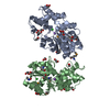


 Mass: 22.990 Da / Num. of mol.: 2 / Source method: obtained synthetically / Formula: Na
 Mass: 35.453 Da / Num. of mol.: 3 / Source method: obtained synthetically / Formula: Cl
 Mass: 59.044 Da / Num. of mol.: 5 / Source method: obtained synthetically / Formula: C2H3O2
 Mass: 62.068 Da / Num. of mol.: 14 / Source method: obtained synthetically / Formula: C2H6O2
 Sample preparation
 / Beamline: BL9-2 / Wavelength: 0.91837, 0.97939
 Processing