 Movie
Movie Controller

[English] 日本語
 Yorodumi
- PDB-2h95: Structure of the Amantadine-Blocked Influenza A M2 Proton Channel... -
+ Open data

- Basic information
| Entry | Database: PDB / ID: 2h95 | ||||||
|---|---|---|---|---|---|---|---|
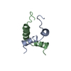
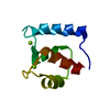
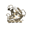
| Title | Structure of the Amantadine-Blocked Influenza A M2 Proton Channel Trans-membrane Domain by Solid-state NMR spectroscopy | ||||||
 Components Components | Matrix protein 2 | ||||||
 Keywords Keywords | MEMBRANE PROTEIN / ALPHA HELIX / PROTEIN-LIGAND | ||||||
| Function / homology |  Function and homology information Function and homology informationsymbiont-mediated suppression of host autophagy / proton transmembrane transporter activity / protein complex oligomerization / host cell membrane / monoatomic ion channel activity / symbiont genome entry into host cell via pore formation in plasma membrane / channel activity / structural constituent of virion / host cell plasma membrane / virion membrane / membrane Similarity search - Function | ||||||
| Method | SOLID-STATE NMR / ENERGY MINIMIZATION WITH ORIENTATIONAL CONSTRAINTS | ||||||
 Authors Authors | Hu, J. / Asbury, T. / Cross, T.A. | ||||||
 Citation Citation | Journal: Biophys.J. / Year: 2007 Title: Backbone structure of the amantadine-blocked trans-membrane domain m2 proton channel from influenza a virus. Authors: Hu, J. / Asbury, T. / Achuthan, S. / Li, C. / Bertram, R. / Quine, J.R. / Fu, R. / Cross, T.A. | ||||||
| History |
|
- Structure visualization
| Structure viewer | Molecule: MolmilJmol/JSmol |
|---|
- Downloads & links
-Download
| PDBx/mmCIF format | 2h95.cif.gz | 28.1 KB | Display | PDBx/mmCIF format |
|---|---|---|---|---|
| PDB format | pdb2h95.ent.gz | 21 KB | Display | PDB format |
| PDBx/mmJSON format | 2h95.json.gz | Tree view | PDBx/mmJSON format | |
| Others |  Other downloads Other downloads |
-Validation report
| Arichive directory | https://data.pdbj.org/pub/pdb/validation_reports/h9/2h95ftp://data.pdbj.org/pub/pdb/validation_reports/h9/2h95 | HTTPS FTP |
|---|
-Related structure data
| Similar structure data |
|---|


-Links
 PDBj
PDBj- Assembly
| Deposited unit | 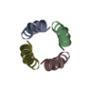
| |||||||||
|---|---|---|---|---|---|---|---|---|---|---|
| 1 |
| |||||||||
| NMR ensembles |
|
-Components
| #1: Protein/peptide | Mass: 1958.496 Da / Num. of mol.: 4 / Fragment: TRANSMEMBRANE DOMAIN (RESIDUES 26-43) / Source method: obtained synthetically Details: THE PEPTIDE WAS SYNTHESIZED USING SOLID PHASE PEPTIDE SYNTHESIS. THIS SEQUENCE OCCURS NATURALLY IN THE INFLUENZA A VIRUS (UDORN/72). References: UniProt: P35938, UniProt: Q3LZC0*PLUS |
|---|
-Experimental details
-Experiment
| Experiment | Method: SOLID-STATE NMR |
|---|---|
| NMR experiment | Type: solid-State NMR PISEMA |
- Sample preparation
| Details | Contents: M2-TMD (~120 mg) and DMPC (~75 mg) were first co-dissolved in 10 ml TFE, followed by the removal of the solvent under vacuum. The peptide/lipid mixture was rehydrated and sonicated to make ...Contents: M2-TMD (~120 mg) and DMPC (~75 mg) were first co-dissolved in 10 ml TFE, followed by the removal of the solvent under vacuum. The peptide/lipid mixture was rehydrated and sonicated to make liposomes in a citrate-borate-phosphate (CBP) buffer (pH 8.8) with 1 mM EDTA and 10 mM amantadine at 310 K. The liposomes were pelleted by ultracentrifugation . Then the pellet was spread on glass slides and dehydrated in a 75% humidity chamber. The dehydrated slides were rehydrated with 1.5 microl liter CBP buffer per slide followed by being stacked into a glass tube and incubated at 316 K for 24 hours in 96% relative humidity. Finally, the glass tube was sealed at both ends with epoxy and two glasscaps. Solvent system: oriented peptide/lipid bilayer of M2_TMD and DMPC |
|---|---|
| Sample conditions | pH: 8.8 / Pressure: AMBIENT / Temperature: 308 K |
-NMR measurement
| Radiation | Protocol: SINGLE WAVELENGTH / Monochromatic (M) / Laue (L): M |
|---|---|
| Radiation wavelength | Relative weight: 1 |
| NMR spectrometer | Type: Bruker AVANCE / Manufacturer: Bruker / Model: AVANCE / Field strength: 400 MHz |
- Processing
| NMR software | Name:  XPLOR-NIH / Version: 2.9.9 / Developer: Schwieters, Kuszewski, Tjandra, Clore / Classification: refinement XPLOR-NIH / Version: 2.9.9 / Developer: Schwieters, Kuszewski, Tjandra, Clore / Classification: refinement |
|---|---|
| Refinement | Method: ENERGY MINIMIZATION WITH ORIENTATIONAL CONSTRAINTS / Software ordinal: 1 Details: REFINEMENT WAS CARRIED OUT IN VACUO ON INITIAL MONOMER COORDINATES CONSISTING OF TWO ALPHA-HELICAL FRAGMENTS (3.6 RESIDUES PER TURN) HAVING TILT AND ROTATIONAL ORIENTATIONS WITH RESPECT TO ...Details: REFINEMENT WAS CARRIED OUT IN VACUO ON INITIAL MONOMER COORDINATES CONSISTING OF TWO ALPHA-HELICAL FRAGMENTS (3.6 RESIDUES PER TURN) HAVING TILT AND ROTATIONAL ORIENTATIONS WITH RESPECT TO THE BILAYER DERIVED FROM PISEMA DIPOLAR WAVE ANALYSIS. ENERGY MINIMIZATION USED A GLOBAL PENALTY FUNCTION INCORPORATING ORIENTATIONAL RESTRAINTS, HYDROGEN BONDING AND THE CHARMM EMPIRICAL FUNCTION. THE ORIENTATIONAL RESTRAINTS IMPOSED ON THE STRUCTURE DURING REFINEMENT ARE 16 15N CHEMICAL SHIFTS AND 16 15N-1H DIPOLAR COUPLINGS FROM PISEMA EXPERIMENTS. A SYMMETRIC, TETRAMERIC BUNDLE MODEL OF M2-TMD WAS CONSTRUCTED BY A SERIES OF RIGID-BODY TRANSFORMATIONS OF THE REFINED M2-TMD MONOMER. THE RESULTING HOMO-TETRAMER IS THE LOWEST FREE ENERGY CONFORMER BASED ON ROTATIONAL CONFORMATIONAL SEARCH. NOTE THAT THE HIS37 AND TRP41 SIDECHAIN POSITIONS ARE CONSISTENT WITH MEASURED ORIENTATIONAL CONSTRAINTS. THE ROTAMERIC STATES OF OTHER RESIDUES ARE TAKEN FROM A BACKBONE DEPENDENT SIDECHAIN ROTAMER LIBRARY (SCRWL). |
| NMR representative | Selection criteria: lowest energy |
| NMR ensemble | Conformer selection criteria: structures with the lowest energy Conformers calculated total number: 72 / Conformers submitted total number: 1 |
