 Movie
Movie Controller

[English] 日本語
 Yorodumi
- PDB-2h6h: Y365F Protein Farnesyltransferase Mutant Complexed with a Farnesy... -
+ Open data

- Basic information
| Entry | Database: PDB / ID: 2h6h | |||||||||
|---|---|---|---|---|---|---|---|---|---|---|
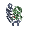
| Title | Y365F Protein Farnesyltransferase Mutant Complexed with a Farnesylated DDPTASACVLS Peptide Product at 1.8A | |||||||||
 Components Components |
| |||||||||
 Keywords Keywords | TRANSFERASE / FTase / farnesyltransferase / farnesyl transferase / prenyltransferase / CaaX / Ras / lipid modification / prenylation / substrate selectivity | |||||||||
| Function / homology |  Function and homology information Function and homology informationskeletal muscle acetylcholine-gated channel clustering / protein geranylgeranyltransferase activity / peptide pheromone maturation / protein farnesylation / protein geranylgeranyltransferase type I / CAAX-protein geranylgeranyltransferase activity / CAAX-protein geranylgeranyltransferase complex / protein farnesyltransferase / protein farnesyltransferase activity / protein farnesyltransferase complex ...skeletal muscle acetylcholine-gated channel clustering / protein geranylgeranyltransferase activity / peptide pheromone maturation / protein farnesylation / protein geranylgeranyltransferase type I / CAAX-protein geranylgeranyltransferase activity / CAAX-protein geranylgeranyltransferase complex / protein farnesyltransferase / protein farnesyltransferase activity / protein farnesyltransferase complex / Rab geranylgeranyltransferase activity / protein geranylgeranylation / regulation of microtubule-based movement / positive regulation of skeletal muscle acetylcholine-gated channel clustering / acetyltransferase activator activity / microtubule associated complex / Apoptotic cleavage of cellular proteins / positive regulation of Rac protein signal transduction / alpha-tubulin binding / positive regulation of cell cycle / peptide binding / transforming growth factor beta receptor signaling pathway / receptor tyrosine kinase binding / lipid metabolic process / RAS processing / Inactivation, recovery and regulation of the phototransduction cascade / microtubule binding / molecular adaptor activity / Potential therapeutics for SARS / positive regulation of cell population proliferation / enzyme binding / zinc ion binding / plasma membrane / cytoplasm / cytosol Similarity search - Function | |||||||||
| Biological species |  Homo sapiens (human) Homo sapiens (human) | |||||||||
| Method |  X-RAY DIFFRACTION / SYNCHROTRON / MOLECULAR REPLACEMENT / Resolution: 1.8 Å X-RAY DIFFRACTION / SYNCHROTRON / MOLECULAR REPLACEMENT / Resolution: 1.8 Å | |||||||||
 Authors Authors | Terry, K.L. / Beese, L.S. | |||||||||
 Citation Citation | Journal: Biochemistry / Year: 2006 Title: Conversion of protein farnesyltransferase to a geranylgeranyltransferase. Authors: Terry, K.L. / Casey, P.J. / Beese, L.S. | |||||||||
| History |
|
- Structure visualization
| Structure viewer | Molecule: MolmilJmol/JSmol |
|---|
- Downloads & links
-Download
| PDBx/mmCIF format | 2h6h.cif.gz | 181.9 KB | Display | PDBx/mmCIF format |
|---|---|---|---|---|
| PDB format | pdb2h6h.ent.gz | 138 KB | Display | PDB format |
| PDBx/mmJSON format | 2h6h.json.gz | Tree view | PDBx/mmJSON format | |
| Others |  Other downloads Other downloads |
-Validation report
| Summary document | 2h6h_validation.pdf.gz | 903.2 KB | Display | wwPDB validaton report |
|---|---|---|---|---|
| Full document | 2h6h_full_validation.pdf.gz | 908.1 KB | Display | |
| Data in XML | 2h6h_validation.xml.gz | 34.3 KB | Display | |
| Data in CIF | 2h6h_validation.cif.gz | 52.3 KB | Display | |
| Arichive directory | https://data.pdbj.org/pub/pdb/validation_reports/h6/2h6hftp://data.pdbj.org/pub/pdb/validation_reports/h6/2h6h | HTTPS FTP |
-Related structure data
| Related structure data |  2h6fC  2h6gC  2h6iC  1jcqS S: Starting model for refinement C: citing same article ( |
|---|---|
| Similar structure data |


-Links
 PDBj
PDBj


- Assembly
| Deposited unit | 
| ||||||||
|---|---|---|---|---|---|---|---|---|---|
| 1 |
| ||||||||
| Unit cell |
|
-Components
-Protein , 2 types, 2 molecules AB
| #1: Protein | Mass: 44864.840 Da / Num. of mol.: 1 Source method: isolated from a genetically manipulated source Source: (gene. exp.) Homo sapiens (human) / Gene: Human FTase alpha subunit / Species (production host): Escherichia coli / Production host:  References: UniProt: P49354, protein farnesyltransferase, protein geranylgeranyltransferase type I |
|---|---|
| #2: Protein | Mass: 48806.406 Da / Num. of mol.: 1 / Mutation: Y365F Source method: isolated from a genetically manipulated source Source: (gene. exp.) Homo sapiens (human) / Gene: Human FTase beta subunit / Species (production host): Escherichia coli / Production host: |
-Protein/peptide / Sugars , 2 types, 2 molecules P
| #3: Protein/peptide | Mass: 1078.152 Da / Num. of mol.: 1 Fragment: residues 173-180 of Rap2a, residues187-189 of H-Ras Mutation: C176T, C177A / Source method: obtained synthetically / Details: Chemically synthesized |
|---|---|
| #4: Polysaccharide | beta-D-fructofuranose-(2-1)-alpha-D-glucopyranose / sucrose  Source method: isolated from a genetically manipulated source Details: oligosaccharide with reducing-end-to-reducing-end glycosidic bond References: sucrose |
-Non-polymers , 4 types, 711 molecules 


| #5: Chemical | ChemComp-ACY /  Mass: 60.052 Da / Num. of mol.: 1 / Source method: obtained synthetically / Formula: C2H4O2 Mass: 60.052 Da / Num. of mol.: 1 / Source method: obtained synthetically / Formula: C2H4O2 |
|---|---|
| #6: Chemical | ChemComp-ZN /  Mass: 65.409 Da / Num. of mol.: 1 / Source method: obtained synthetically / Formula: Zn Mass: 65.409 Da / Num. of mol.: 1 / Source method: obtained synthetically / Formula: Zn |
| #7: Chemical | ChemComp-FAR /  Mass: 206.367 Da / Num. of mol.: 1 / Source method: obtained synthetically / Formula: C15H26 Mass: 206.367 Da / Num. of mol.: 1 / Source method: obtained synthetically / Formula: C15H26 |
| #8: Water | ChemComp-HOH / Mass: 18.015 Da / Num. of mol.: 708 / Source method: isolated from a natural source / Formula: H2O |
-Details
| Has protein modification | Y |
|---|
-Experimental details
-Experiment
| Experiment | Method: X-RAY DIFFRACTION / Number of used crystals: 1 |
|---|
- Sample preparation
| Crystal | Density Matthews: 3.35 Å3/Da / Density % sol: 63.26 % |
|---|---|
| Crystal grow | Temperature: 290 K / Method: vapor diffusion, hanging drop / pH: 5.2 Details: 14% PEG 8000, 200 mM ammonium acetate pH 5.2, 20 mM DTT, VAPOR DIFFUSION, HANGING DROP, temperature 290K |
-Data collection
| Diffraction | Mean temperature: 100 K |
|---|---|
| Diffraction source | Source: SYNCHROTRON / Site: NSLS  / Beamline: X25 / Wavelength: 1.067401 Å / Beamline: X25 / Wavelength: 1.067401 Å |
| Detector | Type: ADSC QUANTUM 315 / Detector: CCD / Date: Feb 28, 2005 / Details: platinum-coated 1:1 focusing toroidal mirror |
| Radiation | Protocol: SINGLE WAVELENGTH / Monochromatic (M) / Laue (L): M / Scattering type: x-ray |
| Radiation wavelength | Wavelength: 1.067401 Å / Relative weight: 1 |
| Reflection | Resolution: 1.8→50 Å / Num. all: 109035 / Num. obs: 109005 / % possible obs: 100 % / Observed criterion σ(I): -3 / Redundancy: 9.2 % / Biso Wilson estimate: 17.7 Å2 / Rsym value: 0.089 / Net I/σ(I): 25.5 |
| Reflection shell | Resolution: 1.8→1.86 Å / Redundancy: 9.1 % / Mean I/σ(I) obs: 6.5 / Num. unique all: 10850 / Rsym value: 0.516 / % possible all: 100 |
- Processing
| Software |
| ||||||||||||||||||||
|---|---|---|---|---|---|---|---|---|---|---|---|---|---|---|---|---|---|---|---|---|---|
| Refinement | Method to determine structure: MOLECULAR REPLACEMENT Starting model: 1JCQ Resolution: 1.8→49.59 Å / Isotropic thermal model: restrained / Cross valid method: THROUGHOUT / σ(F): 0 / Stereochemistry target values: Engh & Huber
| ||||||||||||||||||||
| Displacement parameters | Biso mean: 20.4 Å2
| ||||||||||||||||||||
| Refine analyze |
| ||||||||||||||||||||
| Refinement step | Cycle: LAST / Resolution: 1.8→49.59 Å
| ||||||||||||||||||||
| Refine LS restraints |
| ||||||||||||||||||||
| LS refinement shell | Resolution: 1.8→1.91 Å / Rfactor Rfree error: 0.007
|
