 Movie
Movie Controller

[English] 日本語
 Yorodumi
- PDB-1qbq: STRUCTURE OF RAT FARNESYL PROTEIN TRANSFERASE COMPLEXED WITH A CV... -
+ Open data

- Basic information
| Entry | Database: PDB / ID: 1qbq | ||||||
|---|---|---|---|---|---|---|---|
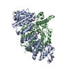
| Title | STRUCTURE OF RAT FARNESYL PROTEIN TRANSFERASE COMPLEXED WITH A CVIM PEPTIDE AND ALPHA-HYDROXYFARNESYLPHOSPHONIC ACID. | ||||||
 Components Components |
| ||||||
 Keywords Keywords | TRANSFERASE / ALPHA-ALPHA-BARREL HELICAL CRESCENT | ||||||
| Function / homology |  Function and homology information Function and homology informationApoptotic cleavage of cellular proteins / Inactivation, recovery and regulation of the phototransduction cascade / RAS processing / protein geranylgeranyltransferase activity / peptide pheromone maturation / protein geranylgeranyltransferase type I / CAAX-protein geranylgeranyltransferase activity / CAAX-protein geranylgeranyltransferase complex / protein farnesylation / protein farnesyltransferase ...Apoptotic cleavage of cellular proteins / Inactivation, recovery and regulation of the phototransduction cascade / RAS processing / protein geranylgeranyltransferase activity / peptide pheromone maturation / protein geranylgeranyltransferase type I / CAAX-protein geranylgeranyltransferase activity / CAAX-protein geranylgeranyltransferase complex / protein farnesylation / protein farnesyltransferase / protein farnesyltransferase activity / protein farnesyltransferase complex / Rab geranylgeranyltransferase activity / protein geranylgeranylation / regulation of fibroblast proliferation / positive regulation of skeletal muscle acetylcholine-gated channel clustering / nuclear envelope organization / geranylgeranyl diphosphate synthase activity / acetyltransferase activator activity / microtubule associated complex / enzyme-linked receptor protein signaling pathway / regulation of microtubule-based movement / alpha-tubulin binding / positive regulation of Rac protein signal transduction / positive regulation of cell cycle / lipid metabolic process / wound healing / protein maturation / receptor tyrosine kinase binding / positive regulation of fibroblast proliferation / microtubule binding / molecular adaptor activity / negative regulation of cell population proliferation / positive regulation of cell population proliferation / negative regulation of apoptotic process / enzyme binding / zinc ion binding / cytoplasm Similarity search - Function | ||||||
| Biological species |  | ||||||
| Method |  X-RAY DIFFRACTION / Resolution: 2.4 Å X-RAY DIFFRACTION / Resolution: 2.4 Å | ||||||
 Authors Authors | Strickland, C.L. / Windsor, W.T. / Syto, R. / Wang, L. / Bond, R. / Wu, Z. / Schwartz, J. / Le, H.V. / Beese, L.S. / Weber, P.C. | ||||||
 Citation Citation | Journal: Biochemistry / Year: 1998 Title: Crystal structure of farnesyl protein transferase complexed with a CaaX peptide and farnesyl diphosphate analogue Authors: Strickland, C.L. / Windsor, W.T. / Syto, R. / Wang, L. / Bond, R. / Wu, Z. / Schwartz, J. / Le, H.V. / Beese, L.S. / Weber, P.C. #1: Journal: Protein Eng. / Year: 1999Title: Engineering of protein farnesyl transferase for co-crystallization. Expression, purification and kinetic characterization of Beta-subunit C-terminal mutants. Authors: Wu, Z. / Demma, M. / Strickland, C.L. / Syto, R. / Le, H.V. / Weber, P.C. / Windsor, W.T. | ||||||
| History |
|
- Structure visualization
| Structure viewer | Molecule: MolmilJmol/JSmol |
|---|
- Downloads & links
-Download
| PDBx/mmCIF format | 1qbq.cif.gz | 166.4 KB | Display | PDBx/mmCIF format |
|---|---|---|---|---|
| PDB format | pdb1qbq.ent.gz | 129.1 KB | Display | PDB format |
| PDBx/mmJSON format | 1qbq.json.gz | Tree view | PDBx/mmJSON format | |
| Others |  Other downloads Other downloads |
-Validation report
| Arichive directory | https://data.pdbj.org/pub/pdb/validation_reports/qb/1qbqftp://data.pdbj.org/pub/pdb/validation_reports/qb/1qbq | HTTPS FTP |
|---|
-Related structure data
| Related structure data | |
|---|---|
| Similar structure data |


-Links
 PDBj
PDBj

- Assembly
| Deposited unit | 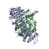
| ||||||||||
|---|---|---|---|---|---|---|---|---|---|---|---|
| 1 |
| ||||||||||
| Unit cell |
|
-Components
-Protein , 2 types, 2 molecules AB
| #1: Protein | Mass: 39760.504 Da / Num. of mol.: 1 Source method: isolated from a genetically manipulated source Details: ALPHA AND BETA SUBUNITS FORM THE BIOLOGICAL UNIT / Source: (gene. exp.) References: UniProt: Q04631, Transferases; Transferring alkyl or aryl groups, other than methyl groups |
|---|---|
| #2: Protein | Mass: 48722.281 Da / Num. of mol.: 1 Source method: isolated from a genetically manipulated source Source: (gene. exp.) References: UniProt: Q02293, Transferases; Transferring alkyl or aryl groups, other than methyl groups |
-Protein/peptide , 1 types, 1 molecules P
| #3: Protein/peptide | Mass: 537.575 Da / Num. of mol.: 1 / Source method: obtained synthetically / Details: Purchased from AnaSpec Inc. |
|---|
-Non-polymers , 4 types, 287 molecules 


| #4: Chemical | ChemComp-ACT /  Mass: 59.044 Da / Num. of mol.: 1 / Source method: obtained synthetically / Formula: C2H3O2 Mass: 59.044 Da / Num. of mol.: 1 / Source method: obtained synthetically / Formula: C2H3O2 |
|---|---|
| #5: Chemical | ChemComp-ZN /  Mass: 65.409 Da / Num. of mol.: 1 / Source method: obtained synthetically / Formula: Zn / Details: Purchased from Calbiochem. Mass: 65.409 Da / Num. of mol.: 1 / Source method: obtained synthetically / Formula: Zn / Details: Purchased from Calbiochem. |
| #6: Chemical | ChemComp-HFP /  Mass: 308.394 Da / Num. of mol.: 1 / Source method: obtained synthetically / Formula: C15H33O4P Mass: 308.394 Da / Num. of mol.: 1 / Source method: obtained synthetically / Formula: C15H33O4P |
| #7: Water | ChemComp-HOH / Mass: 18.015 Da / Num. of mol.: 284 / Source method: isolated from a natural source / Formula: H2O |
-Details
| Has protein modification | Y |
|---|
-Experimental details
-Experiment
| Experiment | Method: X-RAY DIFFRACTION / Number of used crystals: 1 |
|---|
- Sample preparation
| Crystal | Density Matthews: 3.43 Å3/Da / Density % sol: 64.09 % | ||||||||||||||||||||||||||||||||||||||||||||||||||||||
|---|---|---|---|---|---|---|---|---|---|---|---|---|---|---|---|---|---|---|---|---|---|---|---|---|---|---|---|---|---|---|---|---|---|---|---|---|---|---|---|---|---|---|---|---|---|---|---|---|---|---|---|---|---|---|---|
| Crystal grow | Temperature: 295 K / Method: vapor diffusion, hanging drop / pH: 5.7 Details: 7% PEG 4000, 0.1 M sodium acetate, pH 5.7, VAPOR DIFFUSION, HANGING DROP, temperature 295K | ||||||||||||||||||||||||||||||||||||||||||||||||||||||
| Crystal grow | *PLUS Temperature: 22 ℃ / pH: 7.7 | ||||||||||||||||||||||||||||||||||||||||||||||||||||||
| Components of the solutions | *PLUS
|
-Data collection
| Diffraction | Mean temperature: 95 K |
|---|---|
| Diffraction source | Source: ROTATING ANODE / Type: RIGAKU RU200 / Wavelength: 1.5418 |
| Detector | Type: RIGAKU RAXIS IIC / Detector: IMAGE PLATE / Date: Oct 6, 1997 |
| Radiation | Protocol: SINGLE WAVELENGTH / Monochromatic (M) / Laue (L): M / Scattering type: x-ray |
| Radiation wavelength | Wavelength: 1.5418 Å / Relative weight: 1 |
| Reflection | Resolution: 2.4→15 Å / Num. all: 47488 / Num. obs: 42739 / % possible obs: 90 % / Observed criterion σ(F): -3 / Observed criterion σ(I): -3 / Redundancy: 3.3 % / Biso Wilson estimate: 44.2 Å2 / Rmerge(I) obs: 0.061 / Net I/σ(I): 20 |
| Reflection shell | Resolution: 2.4→2.5 Å / Redundancy: 3 % / Rmerge(I) obs: 0.371 / Num. unique all: 47488 / % possible all: 57 |
| Reflection | *PLUS % possible obs: 90 % / Num. measured all: 144714 |
| Reflection shell | *PLUS % possible obs: 57 % / Mean I/σ(I) obs: 3 |
- Processing
| Software |
| ||||||||||||||||||||
|---|---|---|---|---|---|---|---|---|---|---|---|---|---|---|---|---|---|---|---|---|---|
| Refinement | Resolution: 2.4→15 Å / σ(F): 0.5 / σ(I): 1
| ||||||||||||||||||||
| Refinement step | Cycle: LAST / Resolution: 2.4→15 Å
| ||||||||||||||||||||
| Software | *PLUS Name: X-PLOR / Classification: refinement | ||||||||||||||||||||
| Refinement | *PLUS Highest resolution: 2.4 Å / Lowest resolution: 15 Å / σ(F): 0.5 / % reflection Rfree: 5 % / Rfactor obs: 0.218 | ||||||||||||||||||||
| Solvent computation | *PLUS | ||||||||||||||||||||
| Displacement parameters | *PLUS |
