 Movie
Movie Controller

[English] 日本語
 Yorodumi
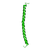
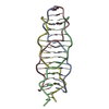
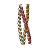
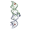
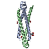
- PDB-2gus: Conformational Transition between Four- and Five-stranded Phenyla... -
+ Open data

- Basic information
| Entry | Database: PDB / ID: 2gus | ||||||
|---|---|---|---|---|---|---|---|
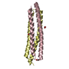
| Title | Conformational Transition between Four- and Five-stranded Phenylalanine Zippers Determined by a Local Packing Interaction | ||||||
 Components Components | Major outer membrane lipoprotein | ||||||
 Keywords Keywords | UNKNOWN FUNCTION / Lipoprotein / protein folding / tetramer / phenylalanine-ZIPPER | ||||||
| Function / homology |  Function and homology information Function and homology informationperiplasmic space organization / lipid modification / peptidoglycan binding / cell outer membrane / outer membrane-bounded periplasmic space / lipid binding / membrane / identical protein binding Similarity search - Function | ||||||
| Biological species |  | ||||||
| Method |  X-RAY DIFFRACTION / SYNCHROTRON / MAD / Resolution: 1.75 Å X-RAY DIFFRACTION / SYNCHROTRON / MAD / Resolution: 1.75 Å | ||||||
 Authors Authors | Liu, J. / Zheng, Q. / Deng, Y. / Kallenbach, N.R. / Lu, M. | ||||||
 Citation Citation | Journal: J.Mol.Biol. / Year: 2006 Title: Conformational Transition between Four and Five-stranded Phenylalanine Zippers Determined by a Local Packing Interaction. Authors: Liu, J. / Zheng, Q. / Deng, Y. / Kallenbach, N.R. / Lu, M. | ||||||
| History |
|
- Structure visualization
| Structure viewer | Molecule: MolmilJmol/JSmol |
|---|
- Downloads & links
-Download
| PDBx/mmCIF format | 2gus.cif.gz | 20.4 KB | Display | PDBx/mmCIF format |
|---|---|---|---|---|
| PDB format | pdb2gus.ent.gz | 12.9 KB | Display | PDB format |
| PDBx/mmJSON format | 2gus.json.gz | Tree view | PDBx/mmJSON format | |
| Others |  Other downloads Other downloads |
-Validation report
| Arichive directory | https://data.pdbj.org/pub/pdb/validation_reports/gu/2gusftp://data.pdbj.org/pub/pdb/validation_reports/gu/2gus | HTTPS FTP |
|---|
-Related structure data


-Links
 PDBj
PDBj


- Assembly
| Deposited unit | 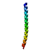
| ||||||||
|---|---|---|---|---|---|---|---|---|---|
| 1 | 
| ||||||||
| Unit cell |
| ||||||||
| Components on special symmetry positions |
| ||||||||
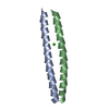
| Details | The biological assembly is a tetramer. |
-Components
| #1: Protein | Mass: 6768.242 Da / Num. of mol.: 1 Mutation: I6F, L9F, V13F, L16F, V20F, L23F, V27M, M30F, V34F, A37F, A41F, A44F, L48F, M51F Source method: isolated from a genetically manipulated source Source: (gene. exp.) |
|---|---|
| #2: Water | ChemComp-HOH /  Mass: 18.015 Da / Num. of mol.: 25 / Source method: isolated from a natural source / Formula: H2O Mass: 18.015 Da / Num. of mol.: 25 / Source method: isolated from a natural source / Formula: H2O |
-Experimental details
-Experiment
| Experiment | Method: X-RAY DIFFRACTION / Number of used crystals: 1 |
|---|
- Sample preparation
| Crystal | Density Matthews: 2.05 Å3/Da / Density % sol: 39.91 % |
|---|---|
| Crystal grow | Temperature: 298 K / Method: vapor diffusion, hanging drop / pH: 8 Details: 0.1M Tris-HCl, 10mM nickel chloride, 14% PEG MME2000, pH 8.0, VAPOR DIFFUSION, HANGING DROP, temperature 298.0K |
-Data collection
| Diffraction | Mean temperature: 100 K | ||||||||||||
|---|---|---|---|---|---|---|---|---|---|---|---|---|---|
| Diffraction source | Source: SYNCHROTRON / Site: NSLS  / Beamline: X4A / Wavelength: 0.9793, 0.9796, 0.9681 / Beamline: X4A / Wavelength: 0.9793, 0.9796, 0.9681 | ||||||||||||
| Detector | Type: ADSC QUANTUM 4 / Detector: CCD / Date: Nov 21, 2005 | ||||||||||||
| Radiation | Monochromator: GRAPHITE / Protocol: MAD / Monochromatic (M) / Laue (L): M / Scattering type: x-ray | ||||||||||||
| Radiation wavelength |
| ||||||||||||
| Reflection | Resolution: 1.75→80.85 Å / Num. all: 6157 / Num. obs: 6157 / % possible obs: 99.1 % / Observed criterion σ(F): 0 / Observed criterion σ(I): 0 / Redundancy: 7.9 % / Biso Wilson estimate: 27.5 Å2 / Rmerge(I) obs: 0.05 / Rsym value: 0.05 / Net I/σ(I): 17 | ||||||||||||
| Reflection shell | Resolution: 1.75→1.81 Å / Redundancy: 7.2 % / Rmerge(I) obs: 0.404 / Mean I/σ(I) obs: 4.4 / Num. unique all: 578 / Rsym value: 0.404 / % possible all: 98.3 |
- Processing
| Software |
| ||||||||||||||||||||||||||||||||||||||||||||||||||||||||||||||||||||||||||||||||||||||||||
|---|---|---|---|---|---|---|---|---|---|---|---|---|---|---|---|---|---|---|---|---|---|---|---|---|---|---|---|---|---|---|---|---|---|---|---|---|---|---|---|---|---|---|---|---|---|---|---|---|---|---|---|---|---|---|---|---|---|---|---|---|---|---|---|---|---|---|---|---|---|---|---|---|---|---|---|---|---|---|---|---|---|---|---|---|---|---|---|---|---|---|---|
| Refinement | Method to determine structure: MAD / Resolution: 1.75→80.85 Å / Cor.coef. Fo:Fc: 0.923 / Cor.coef. Fo:Fc free: 0.894 / SU B: 4.129 / SU ML: 0.066 / TLS residual ADP flag: LIKELY RESIDUAL / Cross valid method: THROUGHOUT / σ(F): 0 / ESU R: 0.134 / ESU R Free: 0.138 / Stereochemistry target values: MAXIMUM LIKELIHOOD
| ||||||||||||||||||||||||||||||||||||||||||||||||||||||||||||||||||||||||||||||||||||||||||
| Solvent computation | Ion probe radii: 0.8 Å / Shrinkage radii: 0.8 Å / VDW probe radii: 1.2 Å / Solvent model: BABINET MODEL WITH MASK | ||||||||||||||||||||||||||||||||||||||||||||||||||||||||||||||||||||||||||||||||||||||||||
| Displacement parameters | Biso mean: 38.101 Å2
| ||||||||||||||||||||||||||||||||||||||||||||||||||||||||||||||||||||||||||||||||||||||||||
| Refinement step | Cycle: LAST / Resolution: 1.75→80.85 Å
| ||||||||||||||||||||||||||||||||||||||||||||||||||||||||||||||||||||||||||||||||||||||||||
| Refine LS restraints |
| ||||||||||||||||||||||||||||||||||||||||||||||||||||||||||||||||||||||||||||||||||||||||||
| LS refinement shell | Resolution: 1.745→1.791 Å / Total num. of bins used: 20
| ||||||||||||||||||||||||||||||||||||||||||||||||||||||||||||||||||||||||||||||||||||||||||
| Refinement TLS params. | Method: refined / Origin x: 4.369 Å / Origin y: 23.026 Å / Origin z: 7.512 Å
|
