 Movie
Movie Controller

[English] 日本語
 Yorodumi
- PDB-2gsc: Crystal Structure of the Conserved Hypothetical Cytosolic Protein... -
+ Open data

- Basic information
| Entry | Database: PDB / ID: 2gsc | ||||||
|---|---|---|---|---|---|---|---|
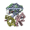
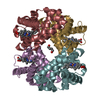
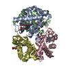
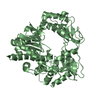
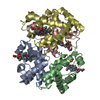
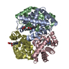
| Title | Crystal Structure of the Conserved Hypothetical Cytosolic Protein Xcc0516 from Xanthomonas campestris | ||||||
 Components Components | Putative uncharacterized protein XCC0516 | ||||||
 Keywords Keywords | UNKNOWN FUNCTION / four-helix bundle / homopentamer / pentameric toroid | ||||||
| Function / homology | 23S rRNA-intervening sequence protein / 23S rRNA-intervening sequence protein / 23S rRNA-intervening sequence / 23S rRNA-intervening sequence superfamily / de novo design (two linked rop proteins) / Up-down Bundle / Mainly Alpha / Four helix bundle protein Function and homology information Function and homology information | ||||||
| Biological species |  Xanthomonas campestris pv. campestris (bacteria) Xanthomonas campestris pv. campestris (bacteria) | ||||||
| Method |  X-RAY DIFFRACTION / SYNCHROTRON / MAD / Resolution: 2.45 Å X-RAY DIFFRACTION / SYNCHROTRON / MAD / Resolution: 2.45 Å | ||||||
 Authors Authors | Lin, L.Y. / Ching, C.L. / Chin, K.H. / Chou, S.H. / Chan, N.L. | ||||||
 Citation Citation | Journal: Proteins / Year: 2006 Title: Crystal structure of the conserved hypothetical cytosolic protein Xcc0516 from Xanthomonas campestris reveals a novel quaternary structure assembled by five four-helix bundles. Authors: Lin, L.Y. / Ching, C.L. / Chin, K.H. / Chou, S.H. / Chan, N.L. | ||||||
| History |
|
- Structure visualization
| Structure viewer | Molecule: MolmilJmol/JSmol |
|---|
- Downloads & links
-Download
| PDBx/mmCIF format | 2gsc.cif.gz | 119.6 KB | Display | PDBx/mmCIF format |
|---|---|---|---|---|
| PDB format | pdb2gsc.ent.gz | 96.7 KB | Display | PDB format |
| PDBx/mmJSON format | 2gsc.json.gz | Tree view | PDBx/mmJSON format | |
| Others |  Other downloads Other downloads |
-Validation report
| Arichive directory | https://data.pdbj.org/pub/pdb/validation_reports/gs/2gscftp://data.pdbj.org/pub/pdb/validation_reports/gs/2gsc | HTTPS FTP |
|---|
-Related structure data
| Similar structure data |
|---|


-Links
 PDBj
PDBj- Assembly
| Deposited unit | 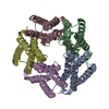
| ||||||||
|---|---|---|---|---|---|---|---|---|---|
| 1 |
| ||||||||
| Unit cell |
| ||||||||
| Components on special symmetry positions |
|
-Components
| #1: Protein | Mass: 14620.450 Da / Num. of mol.: 5 Source method: isolated from a genetically manipulated source Source: (gene. exp.) Xanthomonas campestris pv. campestris (bacteria)Strain: ATCC 33913 / Production host: #2: Water | ChemComp-HOH / |  Mass: 18.015 Da / Num. of mol.: 85 / Source method: isolated from a natural source / Formula: H2O Mass: 18.015 Da / Num. of mol.: 85 / Source method: isolated from a natural source / Formula: H2O |
|---|
-Experimental details
-Experiment
| Experiment | Method: X-RAY DIFFRACTION / Number of used crystals: 1 |
|---|
- Sample preparation
| Crystal | Density Matthews: 2.08 Å3/Da / Density % sol: 40.88 % |
|---|---|
| Crystal grow | Temperature: 277 K / Method: vapor diffusion, hanging drop / pH: 6 Details: 200mM KCl, 5mM MgCl2, 50mM Na cacodylate pH 6.0, 16%(w/v) 1,6-hexanediol, 100mM CsCl, VAPOR DIFFUSION, HANGING DROP, temperature 277K |
-Data collection
| Diffraction | Mean temperature: 100 K | ||||||||||||
|---|---|---|---|---|---|---|---|---|---|---|---|---|---|
| Diffraction source | Source: SYNCHROTRON / Site: SPring-8  / Beamline: BL12B2 / Wavelength: 0.9537, 0.97991, 0.97972 / Beamline: BL12B2 / Wavelength: 0.9537, 0.97991, 0.97972 | ||||||||||||
| Detector | Type: ADSC QUANTUM 4 / Detector: CCD / Date: Jun 20, 2005 | ||||||||||||
| Radiation | Monochromator: double crystal monochromator / Protocol: MAD / Monochromatic (M) / Laue (L): M / Scattering type: x-ray | ||||||||||||
| Radiation wavelength |
| ||||||||||||
| Reflection | Resolution: 2.45→30 Å / Num. all: 23060 / Num. obs: 22715 / % possible obs: 98.5 % / Observed criterion σ(F): 1 / Observed criterion σ(I): 1 / Redundancy: 5.8 % / Rmerge(I) obs: 0.058 / Net I/σ(I): 16.7 | ||||||||||||
| Reflection shell | Resolution: 2.45→2.54 Å / Rmerge(I) obs: 0.146 / % possible all: 98.9 |
- Processing
| Software |
| ||||||||||||||||||||||||||||||||||||||||||||||||||||||||||||||||||||||||||||||||||||||||||
|---|---|---|---|---|---|---|---|---|---|---|---|---|---|---|---|---|---|---|---|---|---|---|---|---|---|---|---|---|---|---|---|---|---|---|---|---|---|---|---|---|---|---|---|---|---|---|---|---|---|---|---|---|---|---|---|---|---|---|---|---|---|---|---|---|---|---|---|---|---|---|---|---|---|---|---|---|---|---|---|---|---|---|---|---|---|---|---|---|---|---|---|
| Refinement | Method to determine structure: MAD / Resolution: 2.45→28.17 Å / Cor.coef. Fo:Fc: 0.912 / Cor.coef. Fo:Fc free: 0.859 / SU B: 9.218 / SU ML: 0.219 / Cross valid method: THROUGHOUT / σ(F): 0 / ESU R: 0.711 / ESU R Free: 0.329 / Stereochemistry target values: MAXIMUM LIKELIHOOD / Details: HYDROGENS HAVE BEEN ADDED IN THE RIDING POSITIONS
| ||||||||||||||||||||||||||||||||||||||||||||||||||||||||||||||||||||||||||||||||||||||||||
| Solvent computation | Ion probe radii: 0.8 Å / Shrinkage radii: 0.8 Å / VDW probe radii: 1.4 Å / Solvent model: MASK | ||||||||||||||||||||||||||||||||||||||||||||||||||||||||||||||||||||||||||||||||||||||||||
| Displacement parameters | Biso mean: 28.148 Å2
| ||||||||||||||||||||||||||||||||||||||||||||||||||||||||||||||||||||||||||||||||||||||||||
| Refinement step | Cycle: LAST / Resolution: 2.45→28.17 Å
| ||||||||||||||||||||||||||||||||||||||||||||||||||||||||||||||||||||||||||||||||||||||||||
| Refine LS restraints |
| ||||||||||||||||||||||||||||||||||||||||||||||||||||||||||||||||||||||||||||||||||||||||||
| LS refinement shell | Resolution: 2.451→2.514 Å / Total num. of bins used: 20
|
