 Movie
Movie Controller

[English] 日本語
 Yorodumi
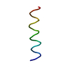
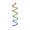
- PDB-2gof: Three-dimensional structure of the trans-membrane domain of Vpu f... -
+ Open data

- Basic information
| Entry | Database: PDB / ID: 2gof | ||||||
|---|---|---|---|---|---|---|---|
| Title | Three-dimensional structure of the trans-membrane domain of Vpu from HIV-1 in aligned phospholipid bicelles | ||||||
 Components Components | VPU protein | ||||||
 Keywords Keywords | VIRAL PROTEIN / single trans-membrane helix / bicelle / magnetic alignment | ||||||
| Function / homology |  Function and homology information Function and homology informationreceptor catabolic process / CD4 receptor binding / viral release from host cell / host cell membrane / monoatomic cation channel activity / symbiont-mediated-mediated suppression of host tetherin activity / symbiont-mediated suppression of host innate immune response / symbiont-mediated suppression of host type I interferon-mediated signaling pathway / membrane Similarity search - Function | ||||||
| Biological species |   Human immunodeficiency virus 1 Human immunodeficiency virus 1 | ||||||
| Method | SOLUTION NMR / Structural fitting | ||||||
| Model type details | minimized average | ||||||
 Authors Authors | Park, S.H. / De Angelis, A.A. / Nevzorov, A.A. / Wu, C.H. / Opella, S.J. | ||||||
 Citation Citation | Journal: Biophys.J. / Year: 2006 Title: Three-Dimensional Structure of the Transmembrane Domain of Vpu from HIV-1 in Aligned Phospholipid Bicelles. Authors: Park, S.H. / De Angelis, A.A. / Nevzorov, A.A. / Wu, C.H. / Opella, S.J. | ||||||
| History |
|
- Structure visualization
| Structure viewer | Molecule: MolmilJmol/JSmol |
|---|
- Downloads & links
-Download
| PDBx/mmCIF format | 2gof.cif.gz | 48.1 KB | Display | PDBx/mmCIF format |
|---|---|---|---|---|
| PDB format | pdb2gof.ent.gz | 24.5 KB | Display | PDB format |
| PDBx/mmJSON format | 2gof.json.gz | Tree view | PDBx/mmJSON format | |
| Others |  Other downloads Other downloads |
-Validation report
| Arichive directory | https://data.pdbj.org/pub/pdb/validation_reports/go/2gofftp://data.pdbj.org/pub/pdb/validation_reports/go/2gof | HTTPS FTP |
|---|
-Related structure data

-Links
 PDBj
PDBj- Assembly
| Deposited unit | 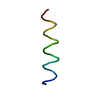
| |||||||||
|---|---|---|---|---|---|---|---|---|---|---|
| 1 |
| |||||||||
| NMR ensembles |
|
-Components
| #1: Protein/peptide | Mass: 3742.689 Da / Num. of mol.: 1 / Fragment: Trans-membrane domain, residues 2-30 / Mutation: Y29G Source method: isolated from a genetically manipulated source Source: (gene. exp.) Human immunodeficiency virus 1 / Genus: Lentivirus / Gene: VPU / Plasmid: pET31-b(+) / Production host:  |
|---|
-Experimental details
-Experiment
| Experiment | Method: SOLUTION NMR |
|---|---|
| NMR experiment | Type: PISEMA |
- Sample preparation
| Details | Type: bicelle Contents: 15N-uniformly or selectively peptide aligned in 16C bicelles (1,2-di-O-tetradecyl-sn-glycero-3-phosphocholine (14-O-PC) / 1,2-di-O-hexyl-sn-glycero-3-phosphocholine (6-O-PC) = 3.0, 28% w/v), 100% H2O Label: sample_1 / Solvent system: 100% H2O |
|---|---|
| Sample conditions | Temperature: 313 K |
-NMR measurement
| Radiation | Protocol: SINGLE WAVELENGTH / Monochromatic (M) / Laue (L): M |
|---|---|
| Radiation wavelength | Relative weight: 1 |
| NMR spectrometer | Type: Bruker AVANCE / Manufacturer: Bruker / Model: AVANCE / Field strength: 900 MHz |
- Processing
| NMR software | Name: Structural Fitting / Developer: Nevzorov, A.A. et al. / Classification: refinement |
|---|---|
| Refinement | Method: Structural fitting / Software ordinal: 1 Details: Orientational frequencies (15N chemical shift and 15N-1H dipolar coupling) for each amide site were used. |
| NMR representative | Selection criteria: minimized average structure |
| NMR ensemble | Conformer selection criteria: first 20 structures / Conformers calculated total number: 100 / Conformers submitted total number: 21 |
