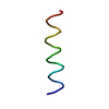
 Movie
Movie Controller

[English] 日本語
 Yorodumi
- PDB-1pje: Structure of the channel-forming trans-membrane domain of Virus p... -
+ Open data

- Basic information
| Entry | Database: PDB / ID: 1pje | ||||||
|---|---|---|---|---|---|---|---|
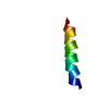
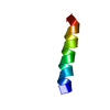
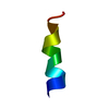
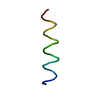
| Title | Structure of the channel-forming trans-membrane domain of Virus protein "u"(Vpu) from HIV-1 | ||||||
 Components Components | VPU protein | ||||||
 Keywords Keywords | VIRAL PROTEIN / ALPHA-HELIX | ||||||
| Function / homology |  Function and homology information Function and homology informationreceptor catabolic process / CD4 receptor binding / viral release from host cell / host cell membrane / monoatomic cation channel activity / symbiont-mediated-mediated suppression of host tetherin activity / symbiont-mediated suppression of host innate immune response / symbiont-mediated suppression of host type I interferon-mediated signaling pathway / membrane Similarity search - Function | ||||||
| Biological species |   Human immunodeficiency virus 1 Human immunodeficiency virus 1 | ||||||
| Method | SOLID-STATE NMR / Direct structural fitting of 2D solid-state NMR data | ||||||
 Authors Authors | Park, S.H. / Mrse, A.A. / Nevzorov, A.A. / Mesleh, M.F. / Oblatt-Montal, M. / Montal, M. / Opella, S.J. | ||||||
 Citation Citation | Journal: J.Mol.Biol. / Year: 2003 Title: Three-dimensional structure of the channel-forming trans-membrane domain of Virus protein "u" (Vpu) from HIV-1 Authors: Park, S.H. / Mrse, A.A. / Nevzorov, A.A. / Mesleh, M.F. / Oblatt-Montal, M. / Montal, M. / Opella, S.J. | ||||||
| History |
|
- Structure visualization
| Structure viewer | Molecule: MolmilJmol/JSmol |
|---|
- Downloads & links
-Download
| PDBx/mmCIF format | 1pje.cif.gz | 11.1 KB | Display | PDBx/mmCIF format |
|---|---|---|---|---|
| PDB format | pdb1pje.ent.gz | 4.2 KB | Display | PDB format |
| PDBx/mmJSON format | 1pje.json.gz | Tree view | PDBx/mmJSON format | |
| Others |  Other downloads Other downloads |
-Validation report
| Arichive directory | https://data.pdbj.org/pub/pdb/validation_reports/pj/1pjeftp://data.pdbj.org/pub/pdb/validation_reports/pj/1pje | HTTPS FTP |
|---|
-Related structure data
-Links
 PDBj
PDBj- Assembly
| Deposited unit | 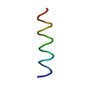
| |||||||||
|---|---|---|---|---|---|---|---|---|---|---|
| 1 |
| |||||||||
| NMR ensembles |
|
-Components
| #1: Protein/peptide | Mass: 3873.885 Da / Num. of mol.: 1 / Fragment: Trans-membrane domain / Mutation: Y29G Source method: isolated from a genetically manipulated source Source: (gene. exp.) Human immunodeficiency virus 1 / Genus: Lentivirus / Gene: VPU / Plasmid: pET-31b(+) / Production host:  |
|---|
-Experimental details
-Experiment
| Experiment | Method: SOLID-STATE NMR |
|---|---|
| NMR experiment | Type: PISEMA |
| NMR details | Text: PISEMA: Polarization Inversion Spin Exchange at the Magic Angle |
- Sample preparation
| Details | Contents: Completely aligned sample in glass plates: 3.5 mg Vpu2-30+ U-15N, 75 mg lipid mixture (DOPC:DOPG, 9:1) |
|---|---|
| Sample conditions | pH: 7 / Pressure: ambient / Temperature: 293 K |
| Crystal grow | *PLUS Method: other / Details: NMR |
-NMR measurement
| Radiation | Protocol: SINGLE WAVELENGTH / Monochromatic (M) / Laue (L): M |
|---|---|
| Radiation wavelength | Relative weight: 1 |
| NMR spectrometer | Type: Bruker AVANCE / Manufacturer: Bruker / Model: AVANCE / Field strength: 700 MHz |
- Processing
| NMR software |
| ||||||||||||||||
|---|---|---|---|---|---|---|---|---|---|---|---|---|---|---|---|---|---|
| Refinement | Method: Direct structural fitting of 2D solid-state NMR data Software ordinal: 1 Details: This structure was calculated by using a structural fitting algorithm that finds torsion angles between consecutive residues based on their NMR frequencies | ||||||||||||||||
| NMR representative | Selection criteria: minimized average structure | ||||||||||||||||
| NMR ensemble | Conformer selection criteria: minimized average structure / Conformers calculated total number: 100 / Conformers submitted total number: 1 |
 NMRPipe