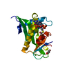
 Movie
Movie Controller


+ Open data

- Basic information
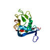
| Entry | Database: PDB / ID: 2go2 | ||||||
|---|---|---|---|---|---|---|---|
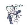
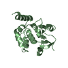
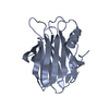
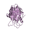
| Title | Crystal structure of BbKI, a Kunitz-type kallikrein inhibitor | ||||||
 Components Components | Kunitz-type serine protease inhibitor BbKI | ||||||
 Keywords Keywords | PROTEIN BINDING / BETA-TREFOIL FOLD | ||||||
| Function / homology |  Function and homology information Function and homology informationendopeptidase inhibitor activity / serine-type endopeptidase inhibitor activity / extracellular region Similarity search - Function | ||||||
| Biological species |  Bauhinia bauhinioides (plant) Bauhinia bauhinioides (plant) | ||||||
| Method |  X-RAY DIFFRACTION / SAD / Resolution: 1.87 Å X-RAY DIFFRACTION / SAD / Resolution: 1.87 Å | ||||||
 Authors Authors | Navarro, M.V.A.S. / Garratt, R.C. | ||||||
 Citation Citation | Journal: To be Published / Year: 2006 Title: The Crystal Structure of BbKI, a kunitz-type kallikrein inhibitor devoid of disulfide bridges Authors: Navarro, M.V.A.S. / Oliva, M.L.V. / Araujo, A.P.U. / Garratt, R.C. #1: Journal: Acta Crystallogr.,Sect.F / Year: 2005 Title: Crystallization and preliminary X-ray analysis of a novel Kunitz-type kallikrein inhibitor from Bauhinia bauhinioides Authors: Navarro, M.V. / Vierira, D.F. / Nagem, R.A. / de Araujo, A.P. / Oliva, M.L. / Garratt, R.C. #2: Journal: Acta Crystallogr.,Sect.D / Year: 2005 Title: Getting the most out of X-ray home sources Authors: Nagem, R.A. / Ambrosio, A.L. / Rojas, A.L. / Navarro, M.V. / Golubev, A.M. / Garratt, R.C. / Polikarpov, I. | ||||||
| History |
|
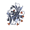
- Structure visualization
| Structure viewer | Molecule: MolmilJmol/JSmol |
|---|
- Downloads & links
-Download
| PDBx/mmCIF format | 2go2.cif.gz | 49 KB | Display | PDBx/mmCIF format |
|---|---|---|---|---|
| PDB format | pdb2go2.ent.gz | 35.7 KB | Display | PDB format |
| PDBx/mmJSON format | 2go2.json.gz | Tree view | PDBx/mmJSON format | |
| Others |  Other downloads Other downloads |
-Validation report
| Arichive directory | https://data.pdbj.org/pub/pdb/validation_reports/go/2go2ftp://data.pdbj.org/pub/pdb/validation_reports/go/2go2 | HTTPS FTP |
|---|
-Related structure data
| Similar structure data |
|---|
-Links
 PDBj
PDBj- Assembly
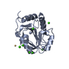
| Deposited unit | 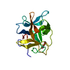
| ||||||||
|---|---|---|---|---|---|---|---|---|---|
| 1 |
| ||||||||
| Unit cell |
| ||||||||
| Details | The asymmetric unit of the crystal contains a monomer, which represents the biological assembly |
-Components
| #1: Protein | Mass: 17839.266 Da / Num. of mol.: 1 Source method: isolated from a genetically manipulated source Source: (gene. exp.) Bauhinia bauhinioides (plant) / Gene: BbKI / Plasmid: pET28 / Species (production host): Escherichia coli / Production host:  | ||
|---|---|---|---|
| #2: Chemical |   Mass: 62.068 Da / Num. of mol.: 2 / Source method: obtained synthetically / Formula: C2H6O2 Mass: 62.068 Da / Num. of mol.: 2 / Source method: obtained synthetically / Formula: C2H6O2#3: Water | ChemComp-HOH / |  Mass: 18.015 Da / Num. of mol.: 160 / Source method: isolated from a natural source / Formula: H2O Mass: 18.015 Da / Num. of mol.: 160 / Source method: isolated from a natural source / Formula: H2O |
-Experimental details
-Experiment
| Experiment | Method: X-RAY DIFFRACTION / Number of used crystals: 1 |
|---|
- Sample preparation
| Crystal | Density Matthews: 2.49 Å3/Da / Density % sol: 50.51 % |
|---|---|
| Crystal grow | Temperature: 293 K / Method: vapor diffusion, hanging drop / pH: 4.6 Details: 8%(w/v) PEG 4000, 0.1 M sodium acetate, pH 4.6, VAPOR DIFFUSION, HANGING DROP, temperature 293K |
-Data collection
| Diffraction | Mean temperature: 100 K |
|---|---|
| Diffraction source | Source: ROTATING ANODE / Type: RIGAKU / Wavelength: 1.5418 Å |
| Detector | Type: MAR scanner 345 mm plate / Detector: IMAGE PLATE / Date: Apr 10, 2004 / Details: mirrors |
| Radiation | Monochromator: GRAPHITE / Protocol: SINGLE WAVELENGTH / Monochromatic (M) / Laue (L): M / Scattering type: x-ray |
| Radiation wavelength | Wavelength: 1.5418 Å / Relative weight: 1 |
| Reflection | Resolution: 1.87→26.44 Å / Num. obs: 14628 / % possible obs: 95.8 % / Observed criterion σ(F): 2 / Observed criterion σ(I): 2 / Redundancy: 8.5 % / Rmerge(I) obs: 0.044 / Net I/σ(I): 32.8 |
| Reflection shell | Resolution: 1.87→1.97 Å / Redundancy: 8.4 % / Rmerge(I) obs: 0.207 / Mean I/σ(I) obs: 10.7 / % possible all: 91.7 |
- Processing
| Software |
| ||||||||||||||||||||||||||||||||||||||||||||||||||||||||||||||||||||||||||||||||||||||||||
|---|---|---|---|---|---|---|---|---|---|---|---|---|---|---|---|---|---|---|---|---|---|---|---|---|---|---|---|---|---|---|---|---|---|---|---|---|---|---|---|---|---|---|---|---|---|---|---|---|---|---|---|---|---|---|---|---|---|---|---|---|---|---|---|---|---|---|---|---|---|---|---|---|---|---|---|---|---|---|---|---|---|---|---|---|---|---|---|---|---|---|---|
| Refinement | Method to determine structure: SAD / Resolution: 1.87→19.73 Å / Cor.coef. Fo:Fc: 0.956 / Cor.coef. Fo:Fc free: 0.944 / SU B: 5.2 / SU ML: 0.083 / TLS residual ADP flag: LIKELY RESIDUAL / Cross valid method: THROUGHOUT / σ(F): 2 / ESU R: 0.138 / ESU R Free: 0.132 / Stereochemistry target values: MAXIMUM LIKELIHOOD / Details: HYDROGENS HAVE BEEN ADDED IN THE RIDING POSITIONS
| ||||||||||||||||||||||||||||||||||||||||||||||||||||||||||||||||||||||||||||||||||||||||||
| Solvent computation | Ion probe radii: 0.8 Å / Shrinkage radii: 0.8 Å / VDW probe radii: 1.4 Å / Solvent model: MASK | ||||||||||||||||||||||||||||||||||||||||||||||||||||||||||||||||||||||||||||||||||||||||||
| Displacement parameters | Biso mean: 14.291 Å2
| ||||||||||||||||||||||||||||||||||||||||||||||||||||||||||||||||||||||||||||||||||||||||||
| Refinement step | Cycle: LAST / Resolution: 1.87→19.73 Å
| ||||||||||||||||||||||||||||||||||||||||||||||||||||||||||||||||||||||||||||||||||||||||||
| Refine LS restraints |
| ||||||||||||||||||||||||||||||||||||||||||||||||||||||||||||||||||||||||||||||||||||||||||
| LS refinement shell | Resolution: 1.87→1.918 Å / Total num. of bins used: 20
| ||||||||||||||||||||||||||||||||||||||||||||||||||||||||||||||||||||||||||||||||||||||||||
| Refinement TLS params. | Method: refined / Origin x: 42.901 Å / Origin y: 58.142 Å / Origin z: 7.866 Å
| ||||||||||||||||||||||||||||||||||||||||||||||||||||||||||||||||||||||||||||||||||||||||||
| Refinement TLS group |
|
