HYDROLASE / Structural genomics / Joint Center for Structural Genomics / JCSG / Protein Structure Initiative / PSI-2
Function / homology
Function and homology information
N-acylneuraminate-9-phosphatase / N-acylneuraminate-9-phosphatase activity / N-acetylglucosamine biosynthetic process / N-acetylneuraminate biosynthetic process / Sialic acid metabolism / CMP-N-acetylneuraminate biosynthetic process / N-acetylneuraminate metabolic process / metal ion binding / cytosol Similarity search - Function
Haloacid dehalogenase hydrolase-like domain / : / HAD-superfamily hydrolase, subfamily IA, CTE7 / Haloacid dehalogenase-like hydrolase / HAD hydrolase, subfamily IA / haloacid dehalogenase-like hydrolase / HAD superfamily/HAD-like / Four Helix Bundle (Hemerythrin (Met), subunit A) / HAD superfamily / HAD-like superfamily ...Haloacid dehalogenase hydrolase-like domain / : / HAD-superfamily hydrolase, subfamily IA, CTE7 / Haloacid dehalogenase-like hydrolase / HAD hydrolase, subfamily IA / haloacid dehalogenase-like hydrolase / HAD superfamily/HAD-like / Four Helix Bundle (Hemerythrin (Met), subunit A) / HAD superfamily / HAD-like superfamily / Up-down Bundle / Rossmann fold / 3-Layer(aba) Sandwich / Mainly Alpha / Alpha Beta Similarity search - Domain/homology
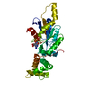
BIOMOLECULE: 1 THIS ENTRY CONTAINS THE CRYSTALLOGRAPHIC ASYMMETRIC UNIT WHICH CONSISTS OF 1 CHAIN(S) ...BIOMOLECULE: 1 THIS ENTRY CONTAINS THE CRYSTALLOGRAPHIC ASYMMETRIC UNIT WHICH CONSISTS OF 1 CHAIN(S). SEE REMARK 350 FOR INFORMATION ON GENERATING THE BIOLOGICAL MOLECULE(S). SIZE EXCLUSION CHROMATOGRAPHY DATA SUPPORTS THE ASSIGNMENT OF A MONOMER AS THE BIOLOGICALLY SIGNIFICANT OLIGOMERIZATION STATE.
Monochromator: Single crystal, cylindrically bent, asymmetrically cut Si(220) crystal Protocol: SINGLE WAVELENGTH / Monochromatic (M) / Laue (L): M / Scattering type: x-ray
Radiation wavelength
Wavelength: 1 Å / Relative weight: 1
Reflection
Resolution: 1.9→50 Å / Num. obs: 24076 / % possible obs: 99.2 % / Redundancy: 8.9 % / Rmerge(I) obs: 0.059 / Χ2: 1.304 / Net I/σ(I): 15.6
Reflection shell
Diffraction-ID: 1
Resolution (Å)
Redundancy (%)
Rmerge(I) obs
Mean I/σ(I) obs
Num. unique obs
Χ2
% possible all
1.9-1.93
5.3
0.47
3.34
1120
1.427
93.8
1.93-1.97
6
0.37
1115
1.445
96
1.97-2.01
6.6
0.325
1166
1.469
99.7
2.01-2.05
8.3
0.294
1191
1.558
100
2.05-2.09
8.6
0.247
1177
1.518
100
2.09-2.14
8.6
0.212
1179
1.47
100
2.14-2.19
8.6
0.198
1184
1.399
100
2.19-2.25
8.5
0.182
1195
1.403
100
2.25-2.32
8.6
0.134
1181
1.308
99.8
2.32-2.39
8.5
0.126
1199
1.282
100
2.39-2.48
8.5
0.111
1193
1.353
100
2.48-2.58
8.5
0.088
1206
1.271
100
2.58-2.7
8.5
0.082
1196
1.358
100
2.7-2.84
9.6
0.081
1211
1.308
99.9
2.84-3.02
11.4
0.073
1214
1.316
99.8
3.02-3.25
11.7
0.061
1232
1.274
99.8
3.25-3.58
11.7
0.053
1230
1.194
99.8
3.58-4.09
11.1
0.045
1241
1.063
99.5
4.09-5.16
10.5
0.046
1279
1.05
99.5
5.16-50
9
0.045
1367
1.07
96.7
-
Phasing
Phasing
Method: molecular replacement
-
Processing
Software
Name
Version
Classification
NB
REFMAC
5.2.0005
refinement
SCALEPACK
datascaling
PDB_EXTRACT
1.701
dataextraction
DENZO
datareduction
MOLREP
phasing
Refinement
Method to determine structure: MOLECULAR REPLACEMENT Starting model: PDb entry 1x42A Resolution: 1.9→44.82 Å / Cor.coef. Fo:Fc: 0.955 / Cor.coef. Fo:Fc free: 0.927 / SU B: 7.019 / SU ML: 0.105 / TLS residual ADP flag: LIKELY RESIDUAL / Cross valid method: THROUGHOUT / σ(F): 0 / ESU R: 0.151 / ESU R Free: 0.151 / Stereochemistry target values: MAXIMUM LIKELIHOOD Details: 1. HYDROGENS HAVE BEEN ADDED IN THE RIDING POSITIONS. 2. NA, CL, PO4, AND EDO MODELED BASED ON CRYSTALIIZATION CONDITIONS AND RELATED STRUCTURES. 3. BASED ON HOMOLOGOUS STRUCTURES, NA COULD ...Details: 1. HYDROGENS HAVE BEEN ADDED IN THE RIDING POSITIONS. 2. NA, CL, PO4, AND EDO MODELED BASED ON CRYSTALIIZATION CONDITIONS AND RELATED STRUCTURES. 3. BASED ON HOMOLOGOUS STRUCTURES, NA COULD ALSO BE MODELED AS MG. 4. THERE IS UNMODELED DENSITY NEAR LYS91 AND LYS243. 5. RESIDUES 63-66 ARE DISORDERED AND WERE NOT MODELED. 6. ATOM RECORD CONTAINS RESIDUAL B FACTORS ONLY.
Rfactor
Num. reflection
% reflection
Selection details
Rfree
0.259
1209
5.1 %
RANDOM
Rwork
0.208
-
-
-
obs
0.21
23709
98.02 %
-
Solvent computation
Ion probe radii: 0.8 Å / Shrinkage radii: 0.8 Å / VDW probe radii: 1.2 Å / Solvent model: MASK
Displacement parameters
Biso mean: 39.234 Å2
Baniso -1
Baniso -2
Baniso -3
1-
0.53 Å2
0.26 Å2
0 Å2
2-
-
0.53 Å2
0 Å2
3-
-
-
-0.79 Å2
Refinement step
Cycle: LAST / Resolution: 1.9→44.82 Å
Protein
Nucleic acid
Ligand
Solvent
Total
Num. atoms
1882
0
11
108
2001
Refine LS restraints
Refine-ID
Type
Dev ideal
Dev ideal target
Number
X-RAY DIFFRACTION
r_bond_refined_d
0.016
0.022
1931
X-RAY DIFFRACTION
r_bond_other_d
0.001
0.02
1780
X-RAY DIFFRACTION
r_angle_refined_deg
1.513
1.97
2613
X-RAY DIFFRACTION
r_angle_other_deg
0.794
3
4127
X-RAY DIFFRACTION
r_dihedral_angle_1_deg
5.536
5
248
X-RAY DIFFRACTION
r_dihedral_angle_2_deg
39.797
24.198
81
X-RAY DIFFRACTION
r_dihedral_angle_3_deg
13.312
15
342
X-RAY DIFFRACTION
r_dihedral_angle_4_deg
13.85
15
13
X-RAY DIFFRACTION
r_chiral_restr
0.085
0.2
304
X-RAY DIFFRACTION
r_gen_planes_refined
0.006
0.02
2138
X-RAY DIFFRACTION
r_gen_planes_other
0.001
0.02
372
X-RAY DIFFRACTION
r_nbd_refined
0.224
0.2
435
X-RAY DIFFRACTION
r_nbd_other
0.171
0.2
1797
X-RAY DIFFRACTION
r_nbtor_refined
0.185
0.2
953
X-RAY DIFFRACTION
r_nbtor_other
0.089
0.2
1205
X-RAY DIFFRACTION
r_xyhbond_nbd_refined
0.163
0.2
107
X-RAY DIFFRACTION
r_metal_ion_refined
0.112
0.2
4
X-RAY DIFFRACTION
r_symmetry_vdw_refined
0.061
0.2
6
X-RAY DIFFRACTION
r_symmetry_vdw_other
0.255
0.2
43
X-RAY DIFFRACTION
r_symmetry_hbond_refined
0.176
0.2
4
X-RAY DIFFRACTION
r_mcbond_it
1.946
3
1226
X-RAY DIFFRACTION
r_mcbond_other
0.581
3
504
X-RAY DIFFRACTION
r_mcangle_it
3.093
5
1969
X-RAY DIFFRACTION
r_scbond_it
5.042
8
723
X-RAY DIFFRACTION
r_scangle_it
7.043
11
642
LS refinement shell
Resolution: 1.897→1.947 Å / Total num. of bins used: 20
Rfactor
Num. reflection
% reflection
Rfree
0.287
87
-
Rwork
0.241
1532
-
obs
-
1619
93.58 %
Refinement TLS params.
Method: refined / Refine-ID: X-RAY DIFFRACTION
ID
L11 (°2)
L12 (°2)
L13 (°2)
L22 (°2)
L23 (°2)
L33 (°2)
S11 (Å °)
S12 (Å °)
S13 (Å °)
S21 (Å °)
S22 (Å °)
S23 (Å °)
S31 (Å °)
S32 (Å °)
S33 (Å °)
T11 (Å2)
T12 (Å2)
T13 (Å2)
T22 (Å2)
T23 (Å2)
T33 (Å2)
Origin x (Å)
Origin y (Å)
Origin z (Å)
1
2.3989
-0.5049
0.5054
2.5462
-0.3751
3.1544
0.1476
-0.1091
0.0969
0.0414
0.056
0.1134
-0.0496
-0.2443
-0.2036
-0.1375
0.0347
0.0839
-0.1115
0.0272
-0.2368
6.205
21.222
11.852
2
2.871
-1.2023
0.9534
3.9727
-1.4669
4.9017
0.0632
0.2413
0.1186
-0.6069
-0.1244
-0.0383
0.1565
0.2451
0.0613
-0.0271
0.0872
-0.0255
0.0108
0.044
-0.0845
-6.06
29.725
-14.221
Refinement TLS group
Refine-ID: X-RAY DIFFRACTION / Selection: ALL / Auth asym-ID: A / Label asym-ID: A
ID
Refine TLS-ID
Auth seq-ID
Label seq-ID
1
1
-2 - 18
10 - 30
2
1
109 - 247
121 - 259
3
2
19 - 108
31 - 120
+
About Yorodumi
-
News
-
Feb 9, 2022. New format data for meta-information of EMDB entries
New format data for meta-information of EMDB entries
Version 3 of the EMDB header file is now the official format.
The previous official version 1.9 will be removed from the archive.
In the structure databanks used in Yorodumi, some data are registered as the other names, "COVID-19 virus" and "2019-nCoV". Here are the details of the virus and the list of structure data.
Jan 31, 2019. EMDB accession codes are about to change! (news from PDBe EMDB page)
EMDB accession codes are about to change! (news from PDBe EMDB page)
The allocation of 4 digits for EMDB accession codes will soon come to an end. Whilst these codes will remain in use, new EMDB accession codes will include an additional digit and will expand incrementally as the available range of codes is exhausted. The current 4-digit format prefixed with “EMD-” (i.e. EMD-XXXX) will advance to a 5-digit format (i.e. EMD-XXXXX), and so on. It is currently estimated that the 4-digit codes will be depleted around Spring 2019, at which point the 5-digit format will come into force.
The EM Navigator/Yorodumi systems omit the EMD- prefix.
Related info.:Q: What is EMD? / ID/Accession-code notation in Yorodumi/EM Navigator
Yorodumi is a browser for structure data from EMDB, PDB, SASBDB, etc.
This page is also the successor to EM Navigator detail page, and also detail information page/front-end page for Omokage search.
The word "yorodu" (or yorozu) is an old Japanese word meaning "ten thousand". "mi" (miru) is to see.
Related info.:EMDB / PDB / SASBDB / Comparison of 3 databanks / Yorodumi Search / Aug 31, 2016. New EM Navigator & Yorodumi / Yorodumi Papers / Jmol/JSmol / Function and homology information / Changes in new EM Navigator and Yorodumi
 Movie
Movie Controller

 Yorodumi
 Open data
 Basic information
 Components
 Keywords
 Function and homology information
 X-RAY DIFFRACTION /
  Authors
 Citation
 Structure visualization
 Downloads & links
 Other downloads


 PDBj
PDBj

 Assembly


 Mass: 22.990 Da / Num. of mol.: 1 / Source method: obtained synthetically / Formula: Na
 Mass: 35.453 Da / Num. of mol.: 1 / Source method: obtained synthetically / Formula: Cl
 Mass: 94.971 Da / Num. of mol.: 1 / Source method: obtained synthetically / Formula: PO4
 Mass: 62.068 Da / Num. of mol.: 1 / Source method: obtained synthetically / Formula: C2H6O2
 Sample preparation
 / Beamline: 5.0.3 / Wavelength: 1
  Processing