 Movie
Movie Controller

[English] 日本語
 Yorodumi
- PDB-2gat: SOLUTION STRUCTURE OF THE C-TERMINAL DOMAIN OF CHICKEN GATA-1 BOU... -
+ Open data

- Basic information
| Entry | Database: PDB / ID: 2gat | ||||||
|---|---|---|---|---|---|---|---|
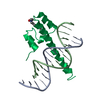
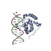
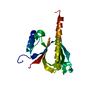
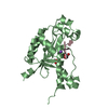
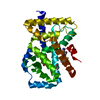
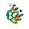
| Title | SOLUTION STRUCTURE OF THE C-TERMINAL DOMAIN OF CHICKEN GATA-1 BOUND TO DNA, NMR, REGULARIZED MEAN STRUCTURE | ||||||
 Components Components |
| ||||||
 Keywords Keywords | TRANSCRIPTION/DNA / DNA BINDING PROTEIN / TRANSCRIPTION FACTOR / ZINC BINDING DOMAIN / COMPLEX (TRANSCRIPTION REGULATION-DNA) / TRANSCRIPTION-DNA COMPLEX | ||||||
| Function / homology |  Function and homology information Function and homology informationcell fate commitment / transcription coactivator binding / DNA-binding transcription factor activity, RNA polymerase II-specific / RNA polymerase II cis-regulatory region sequence-specific DNA binding / negative regulation of transcription by RNA polymerase II / positive regulation of transcription by RNA polymerase II / zinc ion binding / nucleus Similarity search - Function | ||||||
| Biological species |  | ||||||
| Method | SOLUTION NMR / simulated annealing | ||||||
 Authors Authors | Clore, G.M. / Tjandra, N. / Starich, M. / Omichinski, J.G. / Gronenborn, A.M. | ||||||
 Citation Citation | Journal: Nat.Struct.Biol. / Year: 1997 Title: Use of dipolar 1H-15N and 1H-13C couplings in the structure determination of magnetically oriented macromolecules in solution. Authors: Tjandra, N. / Omichinski, J.G. / Gronenborn, A.M. / Clore, G.M. / Bax, A. #1: Journal: J.Mol.Biol. / Year: 1998Title: The solution structure of the Leu22-->Val mutant AREA DNA binding domain complexed with a TGATAG core element defines a role for hydrophobic packing in the determination of specificity Authors: Starich, M.R. / Wikstrom, M. / Shumacher, S. / Arst, H.N. / Gronenborn, A.M. / Clore, G.M. #2: Journal: Science / Year: 1993Title: NMR Structure of a Specific DNA Complex of Zn-Containing DNA Binding Domain of Gata-1 Authors: Omichinski, J.G. / Clore, G.M. / Schaad, O. / Felsenfeld, G. / Trainor, C. / Appella, E. / Stahl, S.J. / Gronenborn, A.M. | ||||||
| History |
|
- Structure visualization
| Structure viewer | Molecule: MolmilJmol/JSmol |
|---|
- Downloads & links
-Download
| PDBx/mmCIF format | 2gat.cif.gz | 63.2 KB | Display | PDBx/mmCIF format |
|---|---|---|---|---|
| PDB format | pdb2gat.ent.gz | 43.5 KB | Display | PDB format |
| PDBx/mmJSON format | 2gat.json.gz | Tree view | PDBx/mmJSON format | |
| Others |  Other downloads Other downloads |
-Validation report
| Arichive directory | https://data.pdbj.org/pub/pdb/validation_reports/ga/2gatftp://data.pdbj.org/pub/pdb/validation_reports/ga/2gat | HTTPS FTP |
|---|
-Related structure data


-Links
 PDBj
PDBj


- Assembly
| Deposited unit | 
| |||||||||
|---|---|---|---|---|---|---|---|---|---|---|
| 1 |
| |||||||||
| NMR ensembles |
|
-Components
| #1: DNA chain | Mass: 4921.228 Da / Num. of mol.: 1 / Source method: obtained synthetically |
|---|---|
| #2: DNA chain | Mass: 4872.190 Da / Num. of mol.: 1 / Source method: obtained synthetically |
| #3: Protein | Mass: 7557.844 Da / Num. of mol.: 1 / Fragment: C-TERMINAL DOMAIN Source method: isolated from a genetically manipulated source Source: (gene. exp.)  |
| #4: Chemical | ChemComp-ZN /   Mass: 65.409 Da / Num. of mol.: 1 / Source method: obtained synthetically / Formula: Zn Mass: 65.409 Da / Num. of mol.: 1 / Source method: obtained synthetically / Formula: Zn |
-Experimental details
-Experiment
| Experiment | Method: SOLUTION NMR |
|---|---|
| NMR details | Text: DATA WERE RECORDED ON A 1:1 COMPLEX OF 1 MOLECULE OF DNA. |
- Sample preparation
| Sample conditions | pH: 6.1 / Temperature: 298 K |
|---|---|
| Crystal grow | *PLUS Method: other / Details: NMR |
-NMR measurement
| NMR spectrometer |
|
|---|
- Processing
| Software |
| ||||||||||||
|---|---|---|---|---|---|---|---|---|---|---|---|---|---|
| NMR software |
| ||||||||||||
| Refinement | Method: simulated annealing / Software ordinal: 1 Details: THE STRUCTURES WERE CALCULATED USING THE SIMULATED ANNEALING PROTOCOL OF NILGES ET AL. (1988) FEBS LETT. 229, 129 - 136 AND PROTEIN ENGINEERING 2, 27 - 38 USING THE PROGRAM X-PLOR MODIFIED ...Details: THE STRUCTURES WERE CALCULATED USING THE SIMULATED ANNEALING PROTOCOL OF NILGES ET AL. (1988) FEBS LETT. 229, 129 - 136 AND PROTEIN ENGINEERING 2, 27 - 38 USING THE PROGRAM X-PLOR MODIFIED TO INCORPORATE DIPOLAR COUPLING RESTRAINTS (TJANDRA ET AL. (1997) NATURE STRUCT BIOL 4, 732-738) AND A CONFORMATIONAL DATABASE POTENTIAL FOR PROTEINS AND NUCLEIC ACIDS (KUSZEWSKI ET AL. (1996) PROTEIN SCI 5, 1067 - 1080 AND (1997) J. MAGN. RESON. 125, 171-177). THE EXPERIMENTAL RESTRAINTS ARE GIVEN IN R2GATMR. THE STRUCTURES ARE BASED ON A TOTAL OF 1830 EXPERIMENTAL NMR RESTRAINTS COMPRISING: 1444 INTERPROTON DISTANCE RESTRAINTS DERIVED FROM NOE MEASUREMENTS; 296 TORSION ANGLE RESTRAINTS; 90 RESIDUAL DIPOLAR COUPLINGS (52 N-H AND 38 C-H). THE NOE RESTRAINTS ARE SUBDIVIDED AS FOLLOWS: (A) WITHIN THE PROTEIN: 242 INTERRESIDUE SEQUENTIAL (|I-J|=1); 161 INTERRESIDUE SHORT RANGE (1(LESS THAN)|I-J|(LESS THAN)=5); 182 INTERRESIDUE LONG RANGE (|I-J|(GREATER THAN)5); AND. 334 INTRARESIDUE. (B) WITHIN THE DNA: 157 INTRARESIDUE; 180 SEQUENTIAL INTRASTRAND; 34 INTERSTRAND; AND 37. H-BONDS (C) BETWEEN PROTEIN AND DNA: 117. THE TORSION ANGLE RESTRAINTS ARE SUBDIVIDED AS FOLLOWS: 144 ANGLES FOR THE PROTEIN (58 PHI, 56 PSI, 26 CHI1 AND 4 CHI2) AND. 152 FOR THE DNA. THE TORSION ANGLE RESTRAINTS FOR THE DNA COMPRISE LOOSE RESTRAINTS ON THE BACKBONE TORSION ANGLES ALPHA, BETA, GAMMA, EPSILON AND ZETA TO PREVENT PROBLEMS ASSOCIATED WITH LOCAL MIRROR IMAGES. THE STRUCTURE IN THIS ENTRY IS THE RESTRAINED REGULARIZED MEAN STRUCTURE AND THE LAST NUMERIC COLUMN REPRESENTS THE RMS OF THE 34 INDIVIDUAL SIMULATED ANNEALING STRUCTURES FOUND IN PDB ENTRY 3GAT ABOUT THE MEAN COORDINATE POSITIONS. THE LAST NUMERIC COLUMN IN THE INDIVIDUAL SA STRUCTURES HAS NO MEANING. THE FOLLOWING TWO SETS OF COORDINATES DEFINE THE PRINCIPAL AXIS OF THE MAGNETIC SUSCEPTIBILITY TENSOR: POINT 1 50.250 -27.763 2.413 POINT 2 50.328 -26.611 3.418 | ||||||||||||
| NMR ensemble | Conformer selection criteria: REGULARIZED MEAN STRUCTURE / Conformers calculated total number: 34 / Conformers submitted total number: 1 |
 X-PLOR