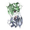
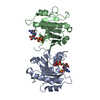
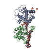
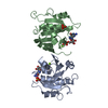
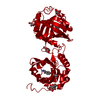
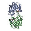
- PDB-2gao: Crystal Structure of Human SAR1a in Complex With GDP -
+
Open data
ID or keywords:
Loading...
-
Basic information
Entry
Database: PDB / ID: 2gao
Title
Crystal Structure of Human SAR1a in Complex With GDP
Components
GTP-binding protein SAR1a
Keywords
PROTEIN TRANSPORT / gtpase / structural genomics consortium / gdp / SGC
Function / homology
Function and homology information
amino acid sensor activity / : / regulation of TORC1 signaling / COPII-coated vesicle cargo loading / cellular response to leucine starvation / COPII vesicle coat assembly / vesicle organization / COPII vesicle coat / membrane organization / Golgi cisterna membrane ...amino acid sensor activity / : / regulation of TORC1 signaling / COPII-coated vesicle cargo loading / cellular response to leucine starvation / COPII vesicle coat assembly / vesicle organization / COPII vesicle coat / membrane organization / Golgi cisterna membrane / endoplasmic reticulum exit site / endoplasmic reticulum to Golgi vesicle-mediated transport / negative regulation of TORC1 signaling / small monomeric GTPase / intracellular protein transport / G protein activity / lysosomal membrane / GTPase activity / endoplasmic reticulum membrane / GTP binding / cytosol Similarity search - Function
Small GTPase SAR1 domain profile. / Small GTPase superfamily, SAR1-type / Sar1p-like members of the Ras-family of small GTPases / Small GTPase superfamily, ARF/SAR type / ADP-ribosylation factor family / ARF-like small GTPases; ARF, ADP-ribosylation factor / Small GTP-binding protein domain / P-loop containing nucleotide triphosphate hydrolases / Rossmann fold / P-loop containing nucleoside triphosphate hydrolase ...Small GTPase SAR1 domain profile. / Small GTPase superfamily, SAR1-type / Sar1p-like members of the Ras-family of small GTPases / Small GTPase superfamily, ARF/SAR type / ADP-ribosylation factor family / ARF-like small GTPases; ARF, ADP-ribosylation factor / Small GTP-binding protein domain / P-loop containing nucleotide triphosphate hydrolases / Rossmann fold / P-loop containing nucleoside triphosphate hydrolase / 3-Layer(aba) Sandwich / Alpha Beta Similarity search - Domain/homology
Resolution: 2→30 Å / Cor.coef. Fo:Fc: 0.927 / Cor.coef. Fo:Fc free: 0.891 / WRfactor Rfree: 0.228 / WRfactor Rwork: 0.191 / SU B: 5.341 / SU ML: 0.146 / ESU R: 0.188 / ESU R Free: 0.175 Details: HYDROGENS HAVE BEEN ADDED IN THE RIDING POSITIONS, ARP/wARP by LAMZIN,PERRAKIS,MORRIS was also used for refinement
Rfactor
Num. reflection
% reflection
Selection details
Rfree
0.2694
1470
5.067 %
thin shells
Rwork
0.2238
-
-
-
all
0.226
-
-
-
obs
-
29010
99.227 %
-
Solvent computation
Ion probe radii: 0.8 Å / Shrinkage radii: 0.8 Å / VDW probe radii: 1.4 Å / Solvent model: MASK BULK SOLVENT
Displacement parameters
Biso mean: 18.669 Å2
Baniso -1
Baniso -2
Baniso -3
1-
0.914 Å2
0 Å2
0.138 Å2
2-
-
-1.376 Å2
0 Å2
3-
-
-
0.538 Å2
Refinement step
Cycle: LAST / Resolution: 2→30 Å
Protein
Nucleic acid
Ligand
Solvent
Total
Num. atoms
2661
0
58
61
2780
Refine LS restraints
Refine-ID
Type
Dev ideal
Dev ideal target
Number
X-RAY DIFFRACTION
r_bond_refined_d
0.017
0.022
2764
X-RAY DIFFRACTION
r_bond_other_d
0.002
0.02
1861
X-RAY DIFFRACTION
r_angle_refined_deg
1.422
2.007
3742
X-RAY DIFFRACTION
r_angle_other_deg
0.953
3
4560
X-RAY DIFFRACTION
r_dihedral_angle_1_deg
4.935
5
334
X-RAY DIFFRACTION
r_dihedral_angle_2_deg
36.112
24.375
112
X-RAY DIFFRACTION
r_dihedral_angle_3_deg
12.564
15
508
X-RAY DIFFRACTION
r_dihedral_angle_4_deg
18.716
15
14
X-RAY DIFFRACTION
r_chiral_restr
0.083
0.2
437
X-RAY DIFFRACTION
r_gen_planes_refined
0.004
0.02
2952
X-RAY DIFFRACTION
r_gen_planes_other
0.001
0.02
542
X-RAY DIFFRACTION
r_nbd_refined
0.2
0.2
491
X-RAY DIFFRACTION
r_nbd_other
0.187
0.2
1917
X-RAY DIFFRACTION
r_nbtor_refined
0.173
0.2
1306
X-RAY DIFFRACTION
r_nbtor_other
0.082
0.2
1327
X-RAY DIFFRACTION
r_xyhbond_nbd_refined
0.156
0.2
91
X-RAY DIFFRACTION
r_symmetry_vdw_refined
0.261
0.2
17
X-RAY DIFFRACTION
r_symmetry_vdw_other
0.205
0.2
41
X-RAY DIFFRACTION
r_symmetry_hbond_refined
0.066
0.2
6
X-RAY DIFFRACTION
r_mcbond_it
2.71
2
2160
X-RAY DIFFRACTION
r_mcbond_other
0.605
2
692
X-RAY DIFFRACTION
r_mcangle_it
3.217
3
2682
X-RAY DIFFRACTION
r_scbond_it
2.218
2
1301
X-RAY DIFFRACTION
r_scangle_it
3.019
3
1058
LS refinement shell
Refine-ID: X-RAY DIFFRACTION / Total num. of bins used: 20
Resolution (Å)
Rfactor Rfree
Num. reflection Rfree
Rfactor Rwork
Num. reflection Rwork
Rfactor all
Num. reflection all
% reflection obs (%)
2-2.052
0.39
98
0.306
1864
0.31
2146
91.426
2.052-2.108
0.336
98
0.293
1936
0.295
2038
99.804
2.108-2.169
0.355
98
0.283
1936
0.286
2036
99.902
2.169-2.235
0.344
98
0.281
1878
0.284
1976
100
2.235-2.308
0.328
98
0.273
1821
0.276
1923
99.792
2.308-2.388
0.301
98
0.25
1784
0.253
1882
100
2.388-2.478
0.29
98
0.236
1681
0.239
1779
100
2.478-2.578
0.27
98
0.234
1616
0.236
1716
99.883
2.578-2.692
0.251
98
0.23
1562
0.231
1660
100
2.692-2.822
0.258
49
0.23
1525
0.231
1575
99.937
2.822-2.973
0.268
98
0.237
1425
0.239
1523
100
2.973-3.152
0.27
49
0.224
1356
0.226
1410
99.645
3.152-3.367
0.278
98
0.208
1253
0.213
1351
100
3.367-3.633
0.236
49
0.191
1200
0.193
1249
100
3.633-3.974
0.259
49
0.181
1112
0.184
1165
99.657
3.974-4.433
0.232
49
0.167
1002
0.17
1053
99.81
4.433-5.1
0.162
49
0.168
885
0.168
935
99.893
5.1-6.202
0.236
49
0.208
742
0.209
791
100
6.202-8.589
0.227
49
0.221
588
0.221
638
99.843
8.589-30
0
0.207
374
0.207
390
95.897
+
About Yorodumi
-
News
-
Feb 9, 2022. New format data for meta-information of EMDB entries
New format data for meta-information of EMDB entries
Version 3 of the EMDB header file is now the official format.
The previous official version 1.9 will be removed from the archive.
In the structure databanks used in Yorodumi, some data are registered as the other names, "COVID-19 virus" and "2019-nCoV". Here are the details of the virus and the list of structure data.
Jan 31, 2019. EMDB accession codes are about to change! (news from PDBe EMDB page)
EMDB accession codes are about to change! (news from PDBe EMDB page)
The allocation of 4 digits for EMDB accession codes will soon come to an end. Whilst these codes will remain in use, new EMDB accession codes will include an additional digit and will expand incrementally as the available range of codes is exhausted. The current 4-digit format prefixed with “EMD-” (i.e. EMD-XXXX) will advance to a 5-digit format (i.e. EMD-XXXXX), and so on. It is currently estimated that the 4-digit codes will be depleted around Spring 2019, at which point the 5-digit format will come into force.
The EM Navigator/Yorodumi systems omit the EMD- prefix.
Related info.:Q: What is EMD? / ID/Accession-code notation in Yorodumi/EM Navigator
Yorodumi is a browser for structure data from EMDB, PDB, SASBDB, etc.
This page is also the successor to EM Navigator detail page, and also detail information page/front-end page for Omokage search.
The word "yorodu" (or yorozu) is an old Japanese word meaning "ten thousand". "mi" (miru) is to see.
Related info.:EMDB / PDB / SASBDB / Comparison of 3 databanks / Yorodumi Search / Aug 31, 2016. New EM Navigator & Yorodumi / Yorodumi Papers / Jmol/JSmol / Function and homology information / Changes in new EM Navigator and Yorodumi
 Movie
Movie Controller


 Open data
 Basic information
 Components
 Keywords
 Function and homology information
 Homo sapiens (human)
 X-RAY DIFFRACTION /
  Authors
 Citation
 Structure visualization
 Downloads & links
 Other downloads
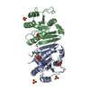


 PDBj
PDBj


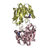
 Assembly


 Type: RNA linking / Mass: 443.201 Da / Num. of mol.: 2 / Source method: obtained synthetically / Formula: C10H15N5O11P2 / Comment: GDP, energy-carrying molecule*YM
 Num. of mol.: 2 / Source method: obtained synthetically
 Mass: 18.015 Da / Num. of mol.: 61 / Source method: isolated from a natural source / Formula: H2O
 Sample preparation
 Processing