- PDB-2fa9: The crystal structure of Sar1[H79G]-GDP provides insight into the... -
+
Open data
ID or keywords:
Loading...
-
Basic information
Entry
Database: PDB / ID: 2fa9
Title
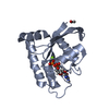
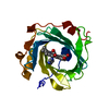
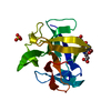
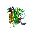
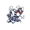
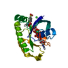
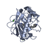
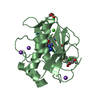
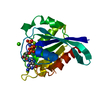
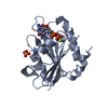
The crystal structure of Sar1[H79G]-GDP provides insight into the coat-controlled GTP hydrolysis in the disassembly of COP II
Components
GTP-binding protein SAR1b
Keywords
PROTEIN TRANSPORT / Sar1H79G mutant
Function / homology
Function and homology information
lipid export from cell / amino acid sensor activity / regulation of lipid transport / lipoprotein transport / COPII-coated vesicle cargo loading / cellular response to leucine starvation / COPII vesicle coat assembly / COPII vesicle coat / Golgi cisterna membrane / lipid homeostasis ...lipid export from cell / amino acid sensor activity / regulation of lipid transport / lipoprotein transport / COPII-coated vesicle cargo loading / cellular response to leucine starvation / COPII vesicle coat assembly / COPII vesicle coat / Golgi cisterna membrane / lipid homeostasis / endoplasmic reticulum exit site / endoplasmic reticulum to Golgi vesicle-mediated transport / negative regulation of TORC1 signaling / small monomeric GTPase / intracellular protein transport / G protein activity / lysosomal membrane / endoplasmic reticulum membrane / GTP binding / metal ion binding / cytosol Similarity search - Function
Small GTPase SAR1 domain profile. / Small GTPase superfamily, SAR1-type / Sar1p-like members of the Ras-family of small GTPases / Small GTPase superfamily, ARF/SAR type / ADP-ribosylation factor family / ARF-like small GTPases; ARF, ADP-ribosylation factor / Small GTP-binding protein domain / P-loop containing nucleotide triphosphate hydrolases / Rossmann fold / P-loop containing nucleoside triphosphate hydrolase ...Small GTPase SAR1 domain profile. / Small GTPase superfamily, SAR1-type / Sar1p-like members of the Ras-family of small GTPases / Small GTPase superfamily, ARF/SAR type / ADP-ribosylation factor family / ARF-like small GTPases; ARF, ADP-ribosylation factor / Small GTP-binding protein domain / P-loop containing nucleotide triphosphate hydrolases / Rossmann fold / P-loop containing nucleoside triphosphate hydrolase / 3-Layer(aba) Sandwich / Alpha Beta Similarity search - Domain/homology
Journal: Chin.J.Struct.Chem. / Year: 2006 Title: Crystal Structure of Sar1[H79G]-GDP Which Provides Insight into the Coat-controlled GTP Hydrolysis in the Disassembly of COP II Authors: Rao, Y. / Yuan, C. / Bian, C. / Hou, X. / Li, Y. / Zhao, G. / Ye, X. / Huang, Z. / Huang, M.
In the structure databanks used in Yorodumi, some data are registered as the other names, "COVID-19 virus" and "2019-nCoV". Here are the details of the virus and the list of structure data.
Jan 31, 2019. EMDB accession codes are about to change! (news from PDBe EMDB page)
EMDB accession codes are about to change! (news from PDBe EMDB page)
The allocation of 4 digits for EMDB accession codes will soon come to an end. Whilst these codes will remain in use, new EMDB accession codes will include an additional digit and will expand incrementally as the available range of codes is exhausted. The current 4-digit format prefixed with “EMD-” (i.e. EMD-XXXX) will advance to a 5-digit format (i.e. EMD-XXXXX), and so on. It is currently estimated that the 4-digit codes will be depleted around Spring 2019, at which point the 5-digit format will come into force.
The EM Navigator/Yorodumi systems omit the EMD- prefix.
Related info.:Q: What is EMD? / ID/Accession-code notation in Yorodumi/EM Navigator
Yorodumi is a browser for structure data from EMDB, PDB, SASBDB, etc.
This page is also the successor to EM Navigator detail page, and also detail information page/front-end page for Omokage search.
The word "yorodu" (or yorozu) is an old Japanese word meaning "ten thousand". "mi" (miru) is to see.
Related info.:EMDB / PDB / SASBDB / Comparison of 3 databanks / Yorodumi Search / Aug 31, 2016. New EM Navigator & Yorodumi / Yorodumi Papers / Jmol/JSmol / Function and homology information / Changes in new EM Navigator and Yorodumi
 Movie
Movie Controller

 Yorodumi
 Open data
 Basic information
 Components
 Keywords
 Function and homology information
 Cricetulus griseus (Chinese hamster)
 X-RAY DIFFRACTION /
  Authors
 Citation
 Structure visualization
 Downloads & links
 Other downloads
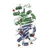

 PDBj
PDBj


 Assembly
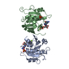


 Mass: 24.305 Da / Num. of mol.: 2 / Source method: obtained synthetically / Formula: Mg
 Mass: 96.063 Da / Num. of mol.: 2 / Source method: obtained synthetically / Formula: SO4
 Type: RNA linking / Mass: 443.201 Da / Num. of mol.: 2 / Source method: obtained synthetically / Formula: C10H15N5O11P2 / Comment: GDP, energy-carrying molecule*YM
 Mass: 18.015 Da / Num. of mol.: 121 / Source method: isolated from a natural source / Formula: H2O
 Sample preparation
 Processing