 Movie
Movie Controller

[English] 日本語
 Yorodumi
- PDB-2gaa: Crystal structure of YFH7 from Saccharomyces cerevisiae: a putati... -
+ Open data

- Basic information
| Entry | Database: PDB / ID: 2gaa | ||||||
|---|---|---|---|---|---|---|---|
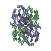
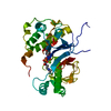
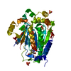
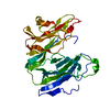
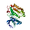
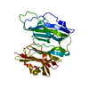
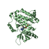
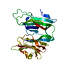
| Title | Crystal structure of YFH7 from Saccharomyces cerevisiae: a putative P-loop containing kinase with a circular permutation. | ||||||
 Components Components | Hypothetical 39.9 kDa protein | ||||||
 Keywords Keywords | UNKNOWN FUNCTION / YFR007w / YFH7 | ||||||
| Function / homology |  Function and homology information Function and homology informationTransferases; Transferring phosphorus-containing groups; Phosphotransferases with an alcohol group as acceptor / phosphorylation / kinase activity / ATP hydrolysis activity / ATP binding / cytoplasm Similarity search - Function | ||||||
| Biological species |  | ||||||
| Method |  X-RAY DIFFRACTION / SYNCHROTRON / FOURIER SYNTHESIS / Resolution: 1.95 Å X-RAY DIFFRACTION / SYNCHROTRON / FOURIER SYNTHESIS / Resolution: 1.95 Å | ||||||
 Authors Authors | Chaptal, V. / Morera, S. | ||||||
 Citation Citation | Journal: Proteins / Year: 2007 Title: Crystal structure and functional analysis identify the P-loop containing protein YFH7 of Saccharomyces cerevisiae as an ATP-dependent kinase. Authors: Gueguen-Chaignon, V. / Chaptal, V. / Lariviere, L. / Costa, N. / Lopes, P. / Morera, S. / Nessler, S. | ||||||
| History |
|
- Structure visualization
| Structure viewer | Molecule: MolmilJmol/JSmol |
|---|
- Downloads & links
-Download
| PDBx/mmCIF format | 2gaa.cif.gz | 82.1 KB | Display | PDBx/mmCIF format |
|---|---|---|---|---|
| PDB format | pdb2gaa.ent.gz | 61.2 KB | Display | PDB format |
| PDBx/mmJSON format | 2gaa.json.gz | Tree view | PDBx/mmJSON format | |
| Others |  Other downloads Other downloads |
-Validation report
| Arichive directory | https://data.pdbj.org/pub/pdb/validation_reports/ga/2gaaftp://data.pdbj.org/pub/pdb/validation_reports/ga/2gaa | HTTPS FTP |
|---|
-Related structure data
| Related structure data |  2ga8SC S: Starting model for refinement C: citing same article ( |
|---|---|
| Similar structure data |


-Links
 PDBj
PDBj- Assembly
| Deposited unit | 
| ||||||||
|---|---|---|---|---|---|---|---|---|---|
| 1 |
| ||||||||
| Unit cell |
|
-Components
| #1: Protein | Mass: 40996.633 Da / Num. of mol.: 1 Source method: isolated from a genetically manipulated source Source: (gene. exp.) Production host:  |
|---|---|
| #2: Chemical | ChemComp-SO4 /   Mass: 96.063 Da / Num. of mol.: 1 / Source method: obtained synthetically / Formula: SO4 Mass: 96.063 Da / Num. of mol.: 1 / Source method: obtained synthetically / Formula: SO4 |
| #3: Water | ChemComp-HOH /  Mass: 18.015 Da / Num. of mol.: 180 / Source method: isolated from a natural source / Formula: H2O Mass: 18.015 Da / Num. of mol.: 180 / Source method: isolated from a natural source / Formula: H2O |
| Has protein modification | Y |
-Experimental details
-Experiment
| Experiment | Method: X-RAY DIFFRACTION / Number of used crystals: 1 |
|---|
- Sample preparation
| Crystal | Density Matthews: 2.07 Å3/Da / Density % sol: 40.7 % |
|---|---|
| Crystal grow | Temperature: 298 K / Method: vapor diffusion, hanging drop / pH: 6.5 Details: PEG 5000MME ammonium sulfate, pH 6.5,VAPOR DIFFUSION, HANGING DROP, temperature 298K |
-Data collection
| Diffraction | Mean temperature: 100 K |
|---|---|
| Diffraction source | Source: SYNCHROTRON / Site: ESRF  / Beamline: ID23-1 / Wavelength: 0.98 Å / Beamline: ID23-1 / Wavelength: 0.98 Å |
| Detector | Type: MARRESEARCH / Detector: CCD / Date: Feb 20, 2005 |
| Radiation | Protocol: SINGLE WAVELENGTH / Monochromatic (M) / Laue (L): M / Scattering type: x-ray |
| Radiation wavelength | Wavelength: 0.98 Å / Relative weight: 1 |
| Reflection | Resolution: 1.95→20 Å / Num. all: 23990 / Num. obs: 23765 / % possible obs: 99.1 % / Observed criterion σ(F): 1 / Observed criterion σ(I): 1 / Redundancy: 2.8 % / Rsym value: 0.059 |
| Reflection shell | Resolution: 1.95→2.06 Å / Redundancy: 2.5 % / Num. unique all: 3436 / Rsym value: 0.378 / % possible all: 99 |
- Processing
| Software |
| ||||||||||||||||||||
|---|---|---|---|---|---|---|---|---|---|---|---|---|---|---|---|---|---|---|---|---|---|
| Refinement | Method to determine structure: FOURIER SYNTHESIS Starting model: 2GA8 Resolution: 1.95→20 Å / σ(F): 2 / Stereochemistry target values: Engh & Huber
| ||||||||||||||||||||
| Refinement step | Cycle: LAST / Resolution: 1.95→20 Å
| ||||||||||||||||||||
| Refine LS restraints |
|
