+ データを開く
- 基本情報
基本情報
| 登録情報 | データベース: PDB / ID: 2g5m | ||||||
|---|---|---|---|---|---|---|---|
| タイトル | Spinophilin PDZ domain | ||||||
 要素 要素 | Neurabin-2 | ||||||
 キーワード キーワード | PROTEIN BINDING / Spinophilin / PDZ domain / CNS / synaptic transmission | ||||||
| 機能・相同性 |  機能・相同性情報 機能・相同性情報protein localization to actin cytoskeleton / positive regulation of protein localization to actin cortical patch / response to L-phenylalanine derivative / protein localization to cell periphery / cytoplasmic side of dendritic spine plasma membrane / cellular response to morphine / spine apparatus / regulation of opioid receptor signaling pathway / developmental process involved in reproduction / extrinsic component of postsynaptic membrane ...protein localization to actin cytoskeleton / positive regulation of protein localization to actin cortical patch / response to L-phenylalanine derivative / protein localization to cell periphery / cytoplasmic side of dendritic spine plasma membrane / cellular response to morphine / spine apparatus / regulation of opioid receptor signaling pathway / developmental process involved in reproduction / extrinsic component of postsynaptic membrane / response to kainic acid / cellular response to peptide / protein phosphatase 1 binding / actin filament depolymerization / filopodium assembly / reproductive system development / dendritic spine membrane / response to steroid hormone / cortical actin cytoskeleton / dendritic spine neck / protein phosphatase inhibitor activity / male mating behavior / dendritic spine head / dendrite development / response to immobilization stress / regulation of postsynapse assembly / response to amino acid / D2 dopamine receptor binding / response to prostaglandin E / positive regulation of protein localization / cellular response to epidermal growth factor stimulus / actin filament organization / response to amphetamine / response to nicotine / learning / adherens junction / positive regulation of protein localization to plasma membrane / hippocampus development / filopodium / cellular response to estradiol stimulus / calcium-mediated signaling / negative regulation of cell growth / cerebral cortex development / kinase binding / ruffle membrane / cellular response to xenobiotic stimulus / neuron projection development / actin filament binding / cell migration / response to estradiol / actin cytoskeleton / lamellipodium / actin binding / growth cone / dendritic spine / transmembrane transporter binding / nucleic acid binding / protein kinase activity / postsynaptic density / response to xenobiotic stimulus / neuronal cell body / dendrite / protein-containing complex binding / glutamatergic synapse / nucleoplasm / plasma membrane / cytoplasm 類似検索 - 分子機能 | ||||||
| 生物種 |  | ||||||
| 手法 | 溶液NMR / simulated annealing torsion angle dynamics | ||||||
 データ登録者 データ登録者 | Kelker, M.S. / Peti, W. | ||||||
 引用 引用 |  ジャーナル: Biochemistry / 年: 2007 ジャーナル: Biochemistry / 年: 2007タイトル: Structural basis for spinophilin-neurabin receptor interaction. 著者: Kelker, M.S. / Dancheck, B. / Ju, T. / Kessler, R.P. / Hudak, J. / Nairn, A.C. / Peti, W. | ||||||
| 履歴 |
|
- 構造の表示
構造の表示
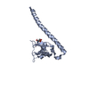
| 構造ビューア | 分子:  Molmil Molmil Jmol/JSmol Jmol/JSmol |
|---|
- ダウンロードとリンク
- ダウンロード
| PDBx/mmCIF形式 |  2g5m.cif.gz 2g5m.cif.gz | 660.7 KB | 表示 |  PDBx/mmCIF形式 PDBx/mmCIF形式 |
|---|---|---|---|---|
| PDB形式 |  pdb2g5m.ent.gz pdb2g5m.ent.gz | 554.9 KB | 表示 |  PDB形式 PDB形式 |
| PDBx/mmJSON形式 |  2g5m.json.gz 2g5m.json.gz | ツリー表示 |  PDBx/mmJSON形式 PDBx/mmJSON形式 | |
| その他 |  その他のダウンロード その他のダウンロード |
-検証レポート
| 文書・要旨 |  2g5m_validation.pdf.gz 2g5m_validation.pdf.gz | 344.6 KB | 表示 |  wwPDB検証レポート wwPDB検証レポート |
|---|---|---|---|---|
| 文書・詳細版 |  2g5m_full_validation.pdf.gz 2g5m_full_validation.pdf.gz | 465.6 KB | 表示 | |
| XML形式データ |  2g5m_validation.xml.gz 2g5m_validation.xml.gz | 33.5 KB | 表示 | |
| CIF形式データ |  2g5m_validation.cif.gz 2g5m_validation.cif.gz | 59.3 KB | 表示 | |
| アーカイブディレクトリ |  https://data.pdbj.org/pub/pdb/validation_reports/g5/2g5m https://data.pdbj.org/pub/pdb/validation_reports/g5/2g5m ftp://data.pdbj.org/pub/pdb/validation_reports/g5/2g5m ftp://data.pdbj.org/pub/pdb/validation_reports/g5/2g5m | HTTPS FTP |
-関連構造データ
| 関連構造データ |  2fn5C C: 同じ文献を引用 ( |
|---|---|
| 類似構造データ | |
| その他のデータベース |
|
- リンク
リンク
- 集合体
集合体
| 登録構造単位 | 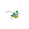
| |||||||||
|---|---|---|---|---|---|---|---|---|---|---|
| 1 |
| |||||||||
| NMR アンサンブル |
|
- 要素
要素
| #1: タンパク質 | 分子量: 12135.745 Da / 分子数: 1 / 断片: PDZ domain / 由来タイプ: 組換発現 / 由来: (組換発現)   |
|---|
-実験情報
-実験
| 実験 | 手法: 溶液NMR | ||||||||||||||||
|---|---|---|---|---|---|---|---|---|---|---|---|---|---|---|---|---|---|
| NMR実験 |
| ||||||||||||||||
| NMR実験の詳細 | Text: The following spectra were used to achieve the sequence-specific backbone and side chain assignments of all aliphatic residues: 2D [1H,15N]-HSQC, 2D [1H,13C]-HSQC, 3D HNCACB, 3D CBCA(CO)NH, 3D ...Text: The following spectra were used to achieve the sequence-specific backbone and side chain assignments of all aliphatic residues: 2D [1H,15N]-HSQC, 2D [1H,13C]-HSQC, 3D HNCACB, 3D CBCA(CO)NH, 3D CC(CO)NH, 3D HNCO, 3D HNCA, 3D HBHA(CO)NH, 3D 15N-resolved [1H,1H]-TOCSY, 3D HC(C)H-TOCSY6. The 2D [1H,1H]-NOESY, 2D [1H,1H]-TOCSY and 2D [1H,1H]-COSY spectra of the Spinophilin493-602 sample in D2O solution after complete H/D exchange of the labile protons were used for the assignment of the aromatic side chains. The NMR spectra were processed with Topspin1.3, and analyzed with the CARA software package. Spectra used for the structure calculation were: 3D 15N-resolved [1H,1H]-NOESY, 3D 13C-resolved [1H,1H]-NOESY (mixing time of 85 ms) and 2D [1H,1H]-NOESY (mixing time 85 ms, D2O solution). |
- 試料調製
試料調製
| 詳細 |
| ||||||||||||
|---|---|---|---|---|---|---|---|---|---|---|---|---|---|
| 試料状態 | イオン強度: 20 mM Na phosphate; 50 mM NaCl / pH: 6.5 / 圧: ambient / 温度: 298 K |
-NMR測定
| 放射 | プロトコル: SINGLE WAVELENGTH / 単色(M)・ラウエ(L): M |
|---|---|
| 放射波長 | 相対比: 1 |
| NMRスペクトロメーター | タイプ: Bruker AVANCE / 製造業者: Bruker / モデル: AVANCE / 磁場強度: 500 MHz |
- 解析
解析
| NMR software |
| ||||||||||||||||||||||||
|---|---|---|---|---|---|---|---|---|---|---|---|---|---|---|---|---|---|---|---|---|---|---|---|---|---|
| 精密化 | 手法: simulated annealing torsion angle dynamics / ソフトェア番号: 1 詳細: The amino acid sequence, the chemical shift assignment, and the NOESY spectra were input for the automated NOESY peak picking and NOE assignment method of ATNOS/CANDID/CYANA. The results of ...詳細: The amino acid sequence, the chemical shift assignment, and the NOESY spectra were input for the automated NOESY peak picking and NOE assignment method of ATNOS/CANDID/CYANA. The results of ATNOS/CANDID/CYANA were refined by manual peak adjustment and additional calculations in CYANA. The 20 conformers from the ATNOS/CANDID cycle 7 with the lowest residual CYANA target function values were energy-minimized in a water shell with the program CNS. | ||||||||||||||||||||||||
| 代表構造 | 選択基準: closest to the average | ||||||||||||||||||||||||
| NMRアンサンブル | コンフォーマー選択の基準: structures with the lowest energy 計算したコンフォーマーの数: 50 / 登録したコンフォーマーの数: 20 |
 ムービー
ムービー コントローラー


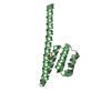
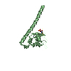
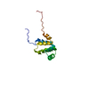
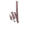
 PDBj
PDBj CYANA
CYANA