 Movie
Movie Controller

[English] 日本語
 Yorodumi
- PDB-2g3p: STRUCTURE OF THE N-TERMINAL TWO DOMAINS OF THE INFECTIVITY PROTEI... -
+ Open data

- Basic information
| Entry | Database: PDB / ID: 2g3p | ||||||
|---|---|---|---|---|---|---|---|
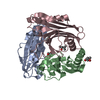
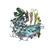
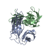
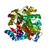
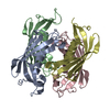
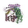
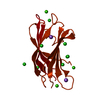
| Title | STRUCTURE OF THE N-TERMINAL TWO DOMAINS OF THE INFECTIVITY PROTEIN G3P OF FILAMENTOUS PHAGE FD | ||||||
 Components Components | INFECTIVITY PROTEIN G3P | ||||||
 Keywords Keywords | VIRAL PROTEIN / INFECTION / FILAMENTOUS PHAGE / PILUS BINDING / BETA BARREL / CONFORMATIONAL CHANGE | ||||||
| Function / homology |  Function and homology information Function and homology informationviral extrusion / virion attachment to host cell pilus / adhesion receptor-mediated virion attachment to host cell / host cell membrane / viral capsid / entry receptor-mediated virion attachment to host cell Similarity search - Function | ||||||
| Biological species |  Enterobacteria phage fd (virus) Enterobacteria phage fd (virus) | ||||||
| Method |  X-RAY DIFFRACTION / SYNCHROTRON / MIRAS / Resolution: 1.9 Å X-RAY DIFFRACTION / SYNCHROTRON / MIRAS / Resolution: 1.9 Å | ||||||
 Authors Authors | Holliger, P. / Williams, R.L. | ||||||
 Citation Citation | Journal: J.Mol.Biol. / Year: 1999 Title: Crystal structure of the two N-terminal domains of g3p from filamentous phage fd at 1.9 A: evidence for conformational lability. Authors: Holliger, P. / Riechmann, L. / Williams, R.L. | ||||||
| History |
|
- Structure visualization
| Structure viewer | Molecule: MolmilJmol/JSmol |
|---|
- Downloads & links
-Download
| PDBx/mmCIF format | 2g3p.cif.gz | 96.5 KB | Display | PDBx/mmCIF format |
|---|---|---|---|---|
| PDB format | pdb2g3p.ent.gz | 73.8 KB | Display | PDB format |
| PDBx/mmJSON format | 2g3p.json.gz | Tree view | PDBx/mmJSON format | |
| Others |  Other downloads Other downloads |
-Validation report
| Arichive directory | https://data.pdbj.org/pub/pdb/validation_reports/g3/2g3pftp://data.pdbj.org/pub/pdb/validation_reports/g3/2g3p | HTTPS FTP |
|---|
-Related structure data
| Similar structure data |
|---|


-Links
 PDBj
PDBj- Assembly
| Deposited unit | 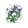
| ||||||||
|---|---|---|---|---|---|---|---|---|---|
| 1 |
| ||||||||
| Unit cell |
| ||||||||
| Noncrystallographic symmetry (NCS) | NCS oper: (Code: given Matrix: (-0.865487, -0.02829, -0.500131), Vector: |
-Components
| #1: Protein | Mass: 24575.518 Da / Num. of mol.: 2 / Fragment: N-TERMINAL 2-DOMAIN FRAGMENT Source method: isolated from a genetically manipulated source Source: (gene. exp.) Enterobacteria phage fd (virus) / Genus: Inovirus / Species: Enterobacteria phage M13 / Plasmid: PUC119 / Cellular location (production host): PERIPLASM / Production host:  #2: Water | ChemComp-HOH / |  Mass: 18.015 Da / Num. of mol.: 303 / Source method: isolated from a natural source / Formula: H2O Mass: 18.015 Da / Num. of mol.: 303 / Source method: isolated from a natural source / Formula: H2OHas protein modification | Y | |
|---|
-Experimental details
-Experiment
| Experiment | Method: X-RAY DIFFRACTION / Number of used crystals: 1 |
|---|
- Sample preparation
| Crystal | Density Matthews: 3.2 Å3/Da / Density % sol: 56 % | ||||||||||||||||||||
|---|---|---|---|---|---|---|---|---|---|---|---|---|---|---|---|---|---|---|---|---|---|
| Crystal grow | pH: 6.2 / Details: pH 6.2 | ||||||||||||||||||||
| Crystal grow | *PLUS Temperature: 21 ℃ / Method: vapor diffusion, hanging drop | ||||||||||||||||||||
| Components of the solutions | *PLUS
|
-Data collection
| Diffraction | Mean temperature: 100 K |
|---|---|
| Diffraction source | Source: SYNCHROTRON / Site: ESRF  / Beamline: ID14-3 / Wavelength: 0.947 / Beamline: ID14-3 / Wavelength: 0.947 |
| Detector | Type: MARRESEARCH / Detector: CCD / Date: Feb 1, 1998 / Details: BENT MIRROR |
| Radiation | Monochromator: SI(111) / Monochromatic (M) / Laue (L): M / Scattering type: x-ray |
| Radiation wavelength | Wavelength: 0.947 Å / Relative weight: 1 |
| Reflection | Resolution: 1.6→13.8 Å / Num. obs: 81474 / % possible obs: 99.7 % / Redundancy: 5.6 % / Biso Wilson estimate: 27 Å2 / Rmerge(I) obs: 0.084 / Rsym value: 0.084 / Net I/σ(I): 4.7 |
| Reflection shell | Resolution: 1.79→1.85 Å / Redundancy: 5.2 % / Rmerge(I) obs: 0.45 / Mean I/σ(I) obs: 1.6 / Rsym value: 0.45 / % possible all: 99.8 |
| Reflection | *PLUS Highest resolution: 1.8 Å / Num. obs: 58927 / % possible obs: 99.8 % / Num. measured all: 343352 / Rmerge(I) obs: 0.079 |
| Reflection shell | *PLUS |
- Processing
| Software |
| ||||||||||||||||||||||||||||||||||||||||||||||||||||||||||||||||||||||||||||||||||||
|---|---|---|---|---|---|---|---|---|---|---|---|---|---|---|---|---|---|---|---|---|---|---|---|---|---|---|---|---|---|---|---|---|---|---|---|---|---|---|---|---|---|---|---|---|---|---|---|---|---|---|---|---|---|---|---|---|---|---|---|---|---|---|---|---|---|---|---|---|---|---|---|---|---|---|---|---|---|---|---|---|---|---|---|---|---|
| Refinement | Method to determine structure: MIRAS / Resolution: 1.9→13 Å / SU B: 3.5 / SU ML: 0.11 / Cross valid method: THROUGHOUT / σ(F): 0 / ESU R: 0.15 / ESU R Free: 0.15
| ||||||||||||||||||||||||||||||||||||||||||||||||||||||||||||||||||||||||||||||||||||
| Displacement parameters | Biso mean: 40 Å2 | ||||||||||||||||||||||||||||||||||||||||||||||||||||||||||||||||||||||||||||||||||||
| Refinement step | Cycle: LAST / Resolution: 1.9→13 Å
| ||||||||||||||||||||||||||||||||||||||||||||||||||||||||||||||||||||||||||||||||||||
| Refine LS restraints |
| ||||||||||||||||||||||||||||||||||||||||||||||||||||||||||||||||||||||||||||||||||||
| Software | *PLUS Name: REFMAC / Classification: refinement | ||||||||||||||||||||||||||||||||||||||||||||||||||||||||||||||||||||||||||||||||||||
| Refinement | *PLUS Rfactor Rfree: 0.299 / Rfactor Rwork: 0.257 | ||||||||||||||||||||||||||||||||||||||||||||||||||||||||||||||||||||||||||||||||||||
| Solvent computation | *PLUS | ||||||||||||||||||||||||||||||||||||||||||||||||||||||||||||||||||||||||||||||||||||
| Displacement parameters | *PLUS | ||||||||||||||||||||||||||||||||||||||||||||||||||||||||||||||||||||||||||||||||||||
| Refine LS restraints | *PLUS
|
