 Movie
Movie Controller

[English] 日本語
 Yorodumi
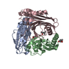
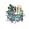
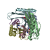
- PDB-2qqd: N47A mutant of Pyruvoyl-dependent Arginine Decarboxylase from Met... -
+ Open data

- Basic information
| Entry | Database: PDB / ID: 2qqd | |||||||||
|---|---|---|---|---|---|---|---|---|---|---|
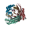
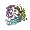
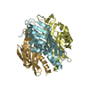
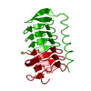
| Title | N47A mutant of Pyruvoyl-dependent Arginine Decarboxylase from Methanococcus jannashii | |||||||||
 Components Components | (Pyruvoyl-dependent arginine decarboxylase (EC 4.1.1.19) ...) x 3 | |||||||||
 Keywords Keywords | LYASE / arginine decarboxylase / pyruvoyl / decarboxylation / autoprocessing / serinolysis / Pyruvate | |||||||||
| Function / homology |  Function and homology information Function and homology informationarginine decarboxylase / arginine decarboxylase activity / L-arginine catabolic process Similarity search - Function | |||||||||
| Biological species |   Methanocaldococcus jannaschii (archaea) Methanocaldococcus jannaschii (archaea) | |||||||||
| Method |  X-RAY DIFFRACTION / SYNCHROTRON / MOLECULAR REPLACEMENT / Resolution: 2 Å X-RAY DIFFRACTION / SYNCHROTRON / MOLECULAR REPLACEMENT / Resolution: 2 Å | |||||||||
 Authors Authors | Ealick, S.E. / Soriano, E.S. | |||||||||
 Citation Citation | Journal: Acta Crystallogr.,Sect.D / Year: 2008 Title: Structures of the N47A and E109Q mutant proteins of pyruvoyl-dependent arginine decarboxylase from Methanococcus jannaschii. Authors: Soriano, E.V. / McCloskey, D.E. / Kinsland, C. / Pegg, A.E. / Ealick, S.E. | |||||||||
| History |
|
- Structure visualization
| Structure viewer | Molecule: MolmilJmol/JSmol |
|---|
- Downloads & links
-Download
| PDBx/mmCIF format | 2qqd.cif.gz | 190 KB | Display | PDBx/mmCIF format |
|---|---|---|---|---|
| PDB format | pdb2qqd.ent.gz | 150.3 KB | Display | PDB format |
| PDBx/mmJSON format | 2qqd.json.gz | Tree view | PDBx/mmJSON format | |
| Others |  Other downloads Other downloads |
-Validation report
| Arichive directory | https://data.pdbj.org/pub/pdb/validation_reports/qq/2qqdftp://data.pdbj.org/pub/pdb/validation_reports/qq/2qqd | HTTPS FTP |
|---|
-Related structure data
| Related structure data |  2qqcC 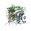 1n13S C: citing same article ( S: Starting model for refinement |
|---|---|
| Similar structure data |


-Links
 PDBj
PDBj

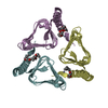
- Assembly
| Deposited unit | 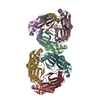
| ||||||||
|---|---|---|---|---|---|---|---|---|---|
| 1 | 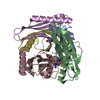
| ||||||||
| 2 | 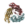
| ||||||||
| Unit cell |
|
-Components
-Pyruvoyl-dependent arginine decarboxylase (EC 4.1.1.19) ... , 3 types, 8 molecules ADBECFGH
| #1: Protein | Mass: 5523.238 Da / Num. of mol.: 2 / Fragment: Beta subunit / Mutation: N47A Source method: isolated from a genetically manipulated source Source: (gene. exp.) Methanocaldococcus jannaschii (archaea)Gene: pdaD / Production host:  #2: Protein | Mass: 12227.369 Da / Num. of mol.: 2 / Fragment: Alpha subunit Source method: isolated from a genetically manipulated source Source: (gene. exp.) Methanocaldococcus jannaschii (archaea)Gene: pdaD / Production host: #3: Protein | Mass: 17819.684 Da / Num. of mol.: 4 / Mutation: N47A Source method: isolated from a genetically manipulated source Source: (gene. exp.) Methanocaldococcus jannaschii (archaea)Gene: pdaD / Production host: |
|---|
-Non-polymers , 4 types, 341 molecules 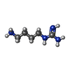


| #4: Chemical |  Mass: 130.191 Da / Num. of mol.: 2 / Source method: obtained synthetically / Formula: C5H14N4 Mass: 130.191 Da / Num. of mol.: 2 / Source method: obtained synthetically / Formula: C5H14N4#5: Chemical |  Mass: 88.062 Da / Num. of mol.: 2 / Source method: obtained synthetically / Formula: C3H4O3 Mass: 88.062 Da / Num. of mol.: 2 / Source method: obtained synthetically / Formula: C3H4O3#6: Chemical |  Mass: 118.174 Da / Num. of mol.: 2 / Source method: obtained synthetically / Formula: C6H14O2 / Comment: precipitant*YM Mass: 118.174 Da / Num. of mol.: 2 / Source method: obtained synthetically / Formula: C6H14O2 / Comment: precipitant*YM#7: Water | ChemComp-HOH / | Mass: 18.015 Da / Num. of mol.: 335 / Source method: isolated from a natural source / Formula: H2O |
|---|
-Experimental details
-Experiment
| Experiment | Method: X-RAY DIFFRACTION / Number of used crystals: 1 |
|---|
- Sample preparation
| Crystal | Density Matthews: 2.14 Å3/Da / Density % sol: 42.49 % |
|---|---|
| Crystal grow | Temperature: 295 K / Method: vapor diffusion, hanging drop / pH: 7 Details: 17-20% PEG 2000, 10% MPD, 2.5% glycerol, 0.1 M HEPES, 0.005 M beta-octylglucoside, 0.005 M EDTA, 0.010 M DTT, pH 7.0, VAPOR DIFFUSION, HANGING DROP, temperature 295K |
-Data collection
| Diffraction | Mean temperature: 100 K |
|---|---|
| Diffraction source | Source: SYNCHROTRON / Site: CHESS  / Beamline: A1 / Wavelength: 0.976 Å / Beamline: A1 / Wavelength: 0.976 Å |
| Detector | Type: ADSC QUANTUM 210 / Detector: CCD / Date: Jun 28, 2004 |
| Radiation | Protocol: SINGLE WAVELENGTH / Monochromatic (M) / Laue (L): M / Scattering type: x-ray |
| Radiation wavelength | Wavelength: 0.976 Å / Relative weight: 1 |
| Reflection | Resolution: 2→46.13 Å / Num. all: 58029 / Num. obs: 56234 / % possible obs: 92.6 % / Observed criterion σ(F): 0 / Observed criterion σ(I): 0 / Redundancy: 4.1 % / Biso Wilson estimate: 14.9 Å2 / Rmerge(I) obs: 0.092 / Rsym value: 0.092 / Net I/σ(I): 16.8 |
| Reflection shell | Resolution: 2→2.13 Å / Redundancy: 2.9 % / Rsym value: 0.04 / % possible all: 83.3 |
- Processing
| Software |
| ||||||||||||||||||||||||||||||||||||
|---|---|---|---|---|---|---|---|---|---|---|---|---|---|---|---|---|---|---|---|---|---|---|---|---|---|---|---|---|---|---|---|---|---|---|---|---|---|
| Refinement | Method to determine structure: MOLECULAR REPLACEMENT Starting model: 1N13 Resolution: 2→46.13 Å / Rfactor Rfree error: 0.003 / Data cutoff high absF: 371512.57 / Data cutoff low absF: 0 / Isotropic thermal model: RESTRAINED / Cross valid method: THROUGHOUT / σ(F): 0 / σ(I): 0 / Stereochemistry target values: Engh & Huber / Details: BULK SOLVENT MODEL USED
| ||||||||||||||||||||||||||||||||||||
| Solvent computation | Solvent model: FLAT MODEL / Bsol: 56.9666 Å2 / ksol: 0.4 e/Å3 | ||||||||||||||||||||||||||||||||||||
| Displacement parameters | Biso mean: 33.7 Å2
| ||||||||||||||||||||||||||||||||||||
| Refine analyze |
| ||||||||||||||||||||||||||||||||||||
| Refinement step | Cycle: LAST / Resolution: 2→46.13 Å
| ||||||||||||||||||||||||||||||||||||
| Refine LS restraints |
| ||||||||||||||||||||||||||||||||||||
| LS refinement shell | Resolution: 2→2.13 Å / Rfactor Rfree error: 0.011 / Total num. of bins used: 6
| ||||||||||||||||||||||||||||||||||||
| Xplor file |
|
