 Movie
Movie Controller

[English] 日本語
 Yorodumi
- PDB-2g2f: A Src-like Inactive Conformation in the Abl Tyrosine Kinase Domain -
+ Open data

- Basic information
| Entry | Database: PDB / ID: 2g2f | ||||||
|---|---|---|---|---|---|---|---|
| Title | A Src-like Inactive Conformation in the Abl Tyrosine Kinase Domain | ||||||
 Components Components |
| ||||||
 Keywords Keywords | TRANSFERASE / protein kinase | ||||||
| Function / homology |  Function and homology information Function and homology informationimmune effector process / protein localization to cytoplasmic microtubule plus-end / mitochondrial depolarization / DNA conformation change / response to epinephrine / phospholipase C-inhibiting G protein-coupled receptor signaling pathway / negative regulation of ubiquitin-protein transferase activity / podocyte apoptotic process / regulation of postsynaptic specialization assembly / positive regulation of phospholipase C/protein kinase C signal transduction ...immune effector process / protein localization to cytoplasmic microtubule plus-end / mitochondrial depolarization / DNA conformation change / response to epinephrine / phospholipase C-inhibiting G protein-coupled receptor signaling pathway / negative regulation of ubiquitin-protein transferase activity / podocyte apoptotic process / regulation of postsynaptic specialization assembly / positive regulation of phospholipase C/protein kinase C signal transduction / regulation of modification of synaptic structure / nicotinate-nucleotide adenylyltransferase activity / delta-catenin binding / Role of ABL in ROBO-SLIT signaling / positive regulation of extracellular matrix organization / neuropilin signaling pathway / neuropilin binding / regulation of cell motility / bubble DNA binding / positive regulation of establishment of T cell polarity / regulation of T cell differentiation / positive regulation of blood vessel branching / proline-rich region binding / cellular response to dopamine / syntaxin binding / positive regulation of dendrite development / regulation of Cdc42 protein signal transduction / regulation of hematopoietic stem cell differentiation / regulation of axon extension / mitogen-activated protein kinase binding / positive regulation of cell migration involved in sprouting angiogenesis / Myogenesis / platelet-derived growth factor receptor-beta signaling pathway / RUNX2 regulates osteoblast differentiation / Fc-gamma receptor signaling pathway involved in phagocytosis / HDR through Single Strand Annealing (SSA) / vascular endothelial cell response to oscillatory fluid shear stress / myoblast proliferation / regulation of endocytosis / associative learning / negative regulation of long-term synaptic potentiation / cardiac muscle cell proliferation / regulation of microtubule polymerization / ephrin receptor signaling pathway / actin monomer binding / cellular response to transforming growth factor beta stimulus / regulation of cell adhesion / positive regulation of focal adhesion assembly / positive regulation of vasoconstriction / positive regulation of substrate adhesion-dependent cell spreading / endothelial cell migration / positive regulation of stress fiber assembly / RHO GTPases Activate WASPs and WAVEs / negative regulation of double-strand break repair via homologous recombination / positive regulation of T cell migration / mismatch repair / ephrin receptor binding / four-way junction DNA binding / ruffle / signal transduction in response to DNA damage / phosphotyrosine residue binding / actin filament polymerization / positive regulation of endothelial cell migration / SH2 domain binding / integrin-mediated signaling pathway / response to endoplasmic reticulum stress / positive regulation of fibroblast proliferation / protein serine/threonine kinase activator activity / Turbulent (oscillatory, disturbed) flow shear stress activates signaling by PIEZO1 and integrins in endothelial cells / regulation of actin cytoskeleton organization / protein kinase C binding / protein modification process / non-specific protein-tyrosine kinase / FCGR3A-mediated phagocytosis / intrinsic apoptotic signaling pathway in response to DNA damage / non-membrane spanning protein tyrosine kinase activity / regulation of autophagy / Regulation of actin dynamics for phagocytic cup formation / kinase activity / cellular response to hydrogen peroxide / epidermal growth factor receptor signaling pathway / enzyme activator activity / positive regulation of neuron apoptotic process / sequence-specific double-stranded DNA binding / actin filament binding / Cyclin D associated events in G1 / actin cytoskeleton / actin cytoskeleton organization / mitotic cell cycle / manganese ion binding / nuclear membrane / Recruitment and ATM-mediated phosphorylation of repair and signaling proteins at DNA double strand breaks / Factors involved in megakaryocyte development and platelet production / MLL4 and MLL3 complexes regulate expression of PPARG target genes in adipogenesis and hepatic steatosis / positive regulation of cytosolic calcium ion concentration / growth cone / RUNX1 regulates transcription of genes involved in differentiation of HSCs / cellular response to oxidative stress / response to oxidative stress / protein tyrosine kinase activity Similarity search - Function | ||||||
| Biological species |  Homo sapiens (human) Homo sapiens (human) | ||||||
| Method |  X-RAY DIFFRACTION / SYNCHROTRON / MOLECULAR REPLACEMENT / Resolution: 2.7 Å X-RAY DIFFRACTION / SYNCHROTRON / MOLECULAR REPLACEMENT / Resolution: 2.7 Å | ||||||
 Authors Authors | Levinson, N.M. / Kuchment, O. | ||||||
 Citation Citation | Journal: Plos Biol. / Year: 2006 Title: A SRC-like inactive conformation in the abl tyrosine kinase domain. Authors: Levinson, N.M. / Kuchment, O. / Shen, K. / Young, M.A. / Koldobskiy, M. / Karplus, M. / Cole, P.A. / Kuriyan, J. | ||||||
| History |
|
- Structure visualization
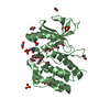
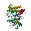
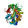
| Structure viewer | Molecule: MolmilJmol/JSmol |
|---|
- Downloads & links
-Download
| PDBx/mmCIF format | 2g2f.cif.gz | 126.2 KB | Display | PDBx/mmCIF format |
|---|---|---|---|---|
| PDB format | pdb2g2f.ent.gz | 97.4 KB | Display | PDB format |
| PDBx/mmJSON format | 2g2f.json.gz | Tree view | PDBx/mmJSON format | |
| Others |  Other downloads Other downloads |
-Validation report
| Arichive directory | https://data.pdbj.org/pub/pdb/validation_reports/g2/2g2fftp://data.pdbj.org/pub/pdb/validation_reports/g2/2g2f | HTTPS FTP |
|---|
-Related structure data
| Related structure data |  2g1tC  2g2hC  2g2iC  1m52S S: Starting model for refinement C: citing same article ( |
|---|---|
| Similar structure data |



-Links
 PDBj
PDBj





- Assembly
| Deposited unit | 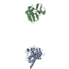
| ||||||||
|---|---|---|---|---|---|---|---|---|---|
| 1 | 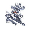
| ||||||||
| 2 | 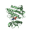
| ||||||||
| Unit cell |
|
-Components
| #1: Protein | Mass: 33181.957 Da / Num. of mol.: 2 / Fragment: Abl Kinase Domain / Mutation: H396P Source method: isolated from a genetically manipulated source Source: (gene. exp.) Homo sapiens (human) / Gene: ABL1, ABL, JTK7 / Production host:   Escherichia coli (E. coli) / References: UniProt: P00519 Escherichia coli (E. coli) / References: UniProt: P00519#2: Protein/peptide | | Mass: 1194.420 Da / Num. of mol.: 1 / Source method: obtained synthetically / Details: solid phase peptide synthesis #3: Chemical | ChemComp-AGS / | 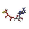 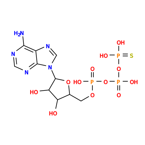 Mass: 523.247 Da / Num. of mol.: 1 / Source method: obtained synthetically / Formula: C10H16N5O12P3S / Comment: ATP-gamma-S, energy-carrying molecule analogue*YM Mass: 523.247 Da / Num. of mol.: 1 / Source method: obtained synthetically / Formula: C10H16N5O12P3S / Comment: ATP-gamma-S, energy-carrying molecule analogue*YM#4: Chemical | ChemComp-112 / | 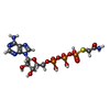 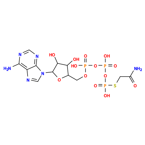 Mass: 580.298 Da / Num. of mol.: 1 / Source method: obtained synthetically / Formula: C12H19N6O13P3S Mass: 580.298 Da / Num. of mol.: 1 / Source method: obtained synthetically / Formula: C12H19N6O13P3S#5: Water | ChemComp-HOH / |  Mass: 18.015 Da / Num. of mol.: 85 / Source method: isolated from a natural source / Formula: H2O Mass: 18.015 Da / Num. of mol.: 85 / Source method: isolated from a natural source / Formula: H2OHas protein modification | Y | |
|---|
-Experimental details
-Experiment
| Experiment | Method: X-RAY DIFFRACTION / Number of used crystals: 1 |
|---|
- Sample preparation
| Crystal | Density Matthews: 2.95 Å3/Da / Density % sol: 58.28 % |
|---|---|
| Crystal grow | Temperature: 291 K / Method: vapor diffusion, hanging drop / pH: 5.5 Details: 0.1M Bis-Tris pH 5.5, 25% PEG 3350, VAPOR DIFFUSION, HANGING DROP, temperature 291K |
-Data collection
| Diffraction | Mean temperature: 100 K |
|---|---|
| Diffraction source | Source: SYNCHROTRON / Site: ALS  / Beamline: 8.2.1 / Wavelength: 1.115879 Å / Beamline: 8.2.1 / Wavelength: 1.115879 Å |
| Detector | Detector: CCD |
| Radiation | Protocol: SINGLE WAVELENGTH / Monochromatic (M) / Laue (L): M / Scattering type: x-ray |
| Radiation wavelength | Wavelength: 1.115879 Å / Relative weight: 1 |
| Reflection | Resolution: 2.7→50 Å / Num. all: 22582 / Num. obs: 22582 / % possible obs: 99.8 % / Observed criterion σ(F): 2 / Observed criterion σ(I): 2 / Rsym value: 0.11 / Net I/σ(I): 10.7 |
| Reflection shell | Resolution: 2.7→2.8 Å / Rmerge(I) obs: 0.494 / Mean I/σ(I) obs: 2.05 / % possible all: 98.2 |
- Processing
| Software |
| ||||||||||||||||||||
|---|---|---|---|---|---|---|---|---|---|---|---|---|---|---|---|---|---|---|---|---|---|
| Refinement | Method to determine structure: MOLECULAR REPLACEMENT Starting model: PDB ENTRY 1M52 Resolution: 2.7→50 Å / Isotropic thermal model: isotropic / σ(F): 0 / Stereochemistry target values: Engh & Huber
| ||||||||||||||||||||
| Displacement parameters | Biso mean: 52.8 Å2 | ||||||||||||||||||||
| Refinement step | Cycle: LAST / Resolution: 2.7→50 Å
| ||||||||||||||||||||
| Refine LS restraints |
|
