 Movie
Movie Controller

[English] 日本語
 Yorodumi
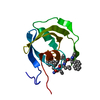
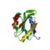
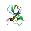
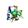
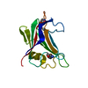
- PDB-2fwg: high resolution crystal structure of the C-terminal domain of the... -
+ Open data

- Basic information
| Entry | Database: PDB / ID: 2fwg | ||||||
|---|---|---|---|---|---|---|---|
| Title | high resolution crystal structure of the C-terminal domain of the electron transfer catalyst DsbD (photoreduced form) | ||||||
 Components Components | Thiol:disulfide interchange protein dsbD | ||||||
 Keywords Keywords | OXIDOREDUCTASE / DSBD / THIOREDOXIN-LIKE / C-terminal domain / photoreduced form | ||||||
| Function / homology |  Function and homology information Function and homology informationprotein-disulfide reductase / protein-disulfide reductase [NAD(P)H] activity / cytochrome complex assembly / response to copper ion / protein-disulfide reductase activity / cell redox homeostasis / electron transfer activity / plasma membrane Similarity search - Function | ||||||
| Biological species |  | ||||||
| Method |  X-RAY DIFFRACTION / SYNCHROTRON / MOLECULAR REPLACEMENT / Resolution: 1.1 Å X-RAY DIFFRACTION / SYNCHROTRON / MOLECULAR REPLACEMENT / Resolution: 1.1 Å | ||||||
 Authors Authors | Stirnimann, C.U. / Rozhkova, A. / Grauschopf, U. / Boeckmann, R.A. / Glockshuber, R. / Capitani, G. / Gruetter, M.G. | ||||||
 Citation Citation | Journal: J.Mol.Biol. / Year: 2006 Title: High-resolution structures of Escherichia coli cDsbD in different redox states: A combined crystallographic, biochemical and computational study Authors: Stirnimann, C.U. / Rozhkova, A. / Grauschopf, U. / Boeckmann, R.A. / Glockshuber, R. / Capitani, G. / Gruetter, M.G. | ||||||
| History |
|
- Structure visualization
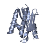
| Structure viewer | Molecule: MolmilJmol/JSmol |
|---|
- Downloads & links
-Download
| PDBx/mmCIF format | 2fwg.cif.gz | 70.9 KB | Display | PDBx/mmCIF format |
|---|---|---|---|---|
| PDB format | pdb2fwg.ent.gz | 51.8 KB | Display | PDB format |
| PDBx/mmJSON format | 2fwg.json.gz | Tree view | PDBx/mmJSON format | |
| Others |  Other downloads Other downloads |
-Validation report
| Arichive directory | https://data.pdbj.org/pub/pdb/validation_reports/fw/2fwgftp://data.pdbj.org/pub/pdb/validation_reports/fw/2fwg | HTTPS FTP |
|---|
-Related structure data
| Related structure data |  2fweC  2fwfC 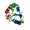 2fwhC  2trxS C: citing same article ( S: Starting model for refinement |
|---|---|
| Similar structure data |


-Links
 PDBj
PDBj

- Assembly
| Deposited unit | 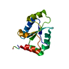
| ||||||||
|---|---|---|---|---|---|---|---|---|---|
| 1 |
| ||||||||
| Unit cell |
|
-Components
| #1: Protein | Mass: 15097.932 Da / Num. of mol.: 1 / Fragment: C-Terminal Domain, Residues 419-546 Source method: isolated from a genetically manipulated source Source: (gene. exp.) |
|---|---|
| #2: Water | ChemComp-HOH /  Mass: 18.015 Da / Num. of mol.: 176 / Source method: isolated from a natural source / Formula: H2O Mass: 18.015 Da / Num. of mol.: 176 / Source method: isolated from a natural source / Formula: H2O |
| Has protein modification | Y |
-Experimental details
-Experiment
| Experiment | Method: X-RAY DIFFRACTION / Number of used crystals: 1 |
|---|
- Sample preparation
| Crystal | Density Matthews: 1.61 Å3/Da / Density % sol: 27.41 % |
|---|---|
| Crystal grow | Temperature: 277.15 K / Method: vapor diffusion, sitting drop / pH: 4.6 Details: 0.1M ammonium acetate, 0.2M sodium acetate, 0.1M sodium iodide, 40% PEG 4000, pH 4.6, VAPOR DIFFUSION, SITTING DROP, temperature 277.15K |
-Data collection
| Diffraction | Mean temperature: 90.9 K |
|---|---|
| Diffraction source | Source: SYNCHROTRON / Site: SLS  / Beamline: X06SA / Wavelength: 0.86 Å / Beamline: X06SA / Wavelength: 0.86 Å |
| Detector | Type: MARRESEARCH / Detector: CCD / Date: May 16, 2003 / Details: dynamically bendable mirror |
| Radiation | Monochromator: Si(111) / Protocol: SINGLE WAVELENGTH / Monochromatic (M) / Laue (L): M / Scattering type: x-ray |
| Radiation wavelength | Wavelength: 0.86 Å / Relative weight: 1 |
| Reflection | Resolution: 1.1→16 Å / Num. all: 42504 / Num. obs: 42360 / % possible obs: 99.7 % / Observed criterion σ(I): -3 / Redundancy: 3.9 % / Biso Wilson estimate: 11.6 Å2 / Rsym value: 0.124 / Net I/σ(I): 6.9 |
| Reflection shell | Resolution: 1.1→1.15 Å / Mean I/σ(I) obs: 3 / Num. unique all: 5200 / Rsym value: 0.429 / % possible all: 99.5 |
- Processing
| Software |
| |||||||||||||||||||||||||||||||||
|---|---|---|---|---|---|---|---|---|---|---|---|---|---|---|---|---|---|---|---|---|---|---|---|---|---|---|---|---|---|---|---|---|---|---|
| Refinement | Method to determine structure: MOLECULAR REPLACEMENT Starting model: modified version of 2TRX (see publication) Resolution: 1.1→16 Å / Num. parameters: 10677 / Num. restraintsaints: 13304 / Cross valid method: THROUGHOUT / σ(F): 0 / Stereochemistry target values: Engh & Huber
| |||||||||||||||||||||||||||||||||
| Refine analyze | Num. disordered residues: 17 / Occupancy sum hydrogen: 893 / Occupancy sum non hydrogen: 1118.2 | |||||||||||||||||||||||||||||||||
| Refinement step | Cycle: LAST / Resolution: 1.1→16 Å
| |||||||||||||||||||||||||||||||||
| Refine LS restraints |
| |||||||||||||||||||||||||||||||||
| LS refinement shell | Highest resolution: 1.1 Å
|
