THIS FILE CONTAINS ONLY A MONOMER, WHILE THE ACTIVE ENZYME IS A DIMER. IN ORDER TO CREATE A DIMERIC MOLECULE, CRYSTALLOGRAPHIC COORDINATES NEED TO BE TRANSFORMED TO (-X), Y, (-Z).
-
Components
#1: Protein
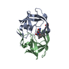
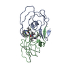
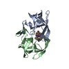
EIAVPROTEASE
Mass: 11392.265 Da / Num. of mol.: 1 / Mutation: I54G Source method: isolated from a genetically manipulated source Source: (gene. exp.) Equine infectious anemia virus / Genus: Lentivirus / Plasmid: PET-226 / Species (production host): Escherichia coli / Production host: Escherichia coli BL21 (bacteria) / Strain (production host): BL21 / References: UniProt: P32542, HIV-1 retropepsin
Method to determine structure: MOLECULAR REPLACEMENT Starting model: HIV-1 PROTEASE Resolution: 1.8→20 Å / σ(F): 3 Details: THE TWO ORIENTATIONS OF THE INHIBITOR CREATED IN THE ACTIVE ENZYME OVERLAP WITH COMPLETE SUPERPOSITION AT THE PERIPHERY AND SOME DEVIATION IN THE CENTER. RESIDUES 56 - 57 HAVE BEEN MODELED ...Details: THE TWO ORIENTATIONS OF THE INHIBITOR CREATED IN THE ACTIVE ENZYME OVERLAP WITH COMPLETE SUPERPOSITION AT THE PERIPHERY AND SOME DEVIATION IN THE CENTER. RESIDUES 56 - 57 HAVE BEEN MODELED WITH TWO ORIENTATIONS OF THEIR MAIN CHAINS, SINCE THEY FORM ALTERNATE HYDROGEN BONDS, DUE TO THE SAME DISORDER PHENOMENON.
Rfactor
Num. reflection
% reflection
Rwork
0.136
-
-
obs
-
8140
85 %
Displacement parameters
Biso mean: 19.12 Å2
Refinement step
Cycle: LAST / Resolution: 1.8→20 Å
Protein
Nucleic acid
Ligand
Solvent
Total
Num. atoms
791
0
40
128
959
Refine LS restraints
Refine-ID
Type
Dev ideal
Dev ideal target
X-RAY DIFFRACTION
p_bond_d
0.014
0.017
X-RAY DIFFRACTION
p_angle_d
0.046
0.044
X-RAY DIFFRACTION
p_angle_deg
X-RAY DIFFRACTION
p_planar_d
0.043
0.059
X-RAY DIFFRACTION
p_hb_or_metal_coord
X-RAY DIFFRACTION
p_mcbond_it
1.916
2.5
X-RAY DIFFRACTION
p_mcangle_it
2.567
3.5
X-RAY DIFFRACTION
p_scbond_it
3.72
4
X-RAY DIFFRACTION
p_scangle_it
5.328
7
X-RAY DIFFRACTION
p_plane_restr
0.018
0.022
X-RAY DIFFRACTION
p_chiral_restr
0.177
0.18
X-RAY DIFFRACTION
p_singtor_nbd
0.207
0.5
X-RAY DIFFRACTION
p_multtor_nbd
0.45
0.5
X-RAY DIFFRACTION
p_xhyhbond_nbd
X-RAY DIFFRACTION
p_xyhbond_nbd
0.382
0.5
X-RAY DIFFRACTION
p_planar_tor
3.4
3.5
X-RAY DIFFRACTION
p_staggered_tor
19.1
14
X-RAY DIFFRACTION
p_orthonormal_tor
X-RAY DIFFRACTION
p_transverse_tor
32.4
12
X-RAY DIFFRACTION
p_special_tor
Software
*PLUS
Name: PROFFT / Classification: refinement
Refinement
*PLUS
Rfactor obs: 0.136
Solvent computation
*PLUS
Displacement parameters
*PLUS
Biso mean: 22.2 Å2
+
About Yorodumi
-
News
-
Feb 9, 2022. New format data for meta-information of EMDB entries
New format data for meta-information of EMDB entries
Version 3 of the EMDB header file is now the official format.
The previous official version 1.9 will be removed from the archive.
In the structure databanks used in Yorodumi, some data are registered as the other names, "COVID-19 virus" and "2019-nCoV". Here are the details of the virus and the list of structure data.
Jan 31, 2019. EMDB accession codes are about to change! (news from PDBe EMDB page)
EMDB accession codes are about to change! (news from PDBe EMDB page)
The allocation of 4 digits for EMDB accession codes will soon come to an end. Whilst these codes will remain in use, new EMDB accession codes will include an additional digit and will expand incrementally as the available range of codes is exhausted. The current 4-digit format prefixed with “EMD-” (i.e. EMD-XXXX) will advance to a 5-digit format (i.e. EMD-XXXXX), and so on. It is currently estimated that the 4-digit codes will be depleted around Spring 2019, at which point the 5-digit format will come into force.
The EM Navigator/Yorodumi systems omit the EMD- prefix.
Related info.:Q: What is EMD? / ID/Accession-code notation in Yorodumi/EM Navigator
Yorodumi is a browser for structure data from EMDB, PDB, SASBDB, etc.
This page is also the successor to EM Navigator detail page, and also detail information page/front-end page for Omokage search.
The word "yorodu" (or yorozu) is an old Japanese word meaning "ten thousand". "mi" (miru) is to see.
Related info.:EMDB / PDB / SASBDB / Comparison of 3 databanks / Yorodumi Search / Aug 31, 2016. New EM Navigator & Yorodumi / Yorodumi Papers / Jmol/JSmol / Function and homology information / Changes in new EM Navigator and Yorodumi
 Movie
Movie Controller


 Open data
 Basic information
 Components
 Keywords
 Function and homology information
 Equine infectious anemia virus
 X-RAY DIFFRACTION /
  Authors
 Citation
 Structure visualization
 Downloads & links
 Other downloads


 PDBj
PDBj
 Assembly
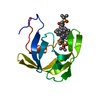


 Mass: 566.751 Da / Num. of mol.: 1 / Source method: obtained synthetically / Formula: C32H42N2O5S
 Mass: 18.015 Da / Num. of mol.: 128 / Source method: isolated from a natural source / Formula: H2O
 Sample preparation
 Processing