 Movie
Movie Controller

[English] 日本語
 Yorodumi
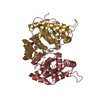
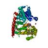
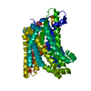
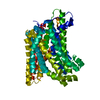
- PDB-2fuj: A putative acyl-CoA thioesterase from Xanthomonas campestris (XC229) -
+ Open data

- Basic information
| Entry | Database: PDB / ID: 2fuj | ||||||
|---|---|---|---|---|---|---|---|
| Title | A putative acyl-CoA thioesterase from Xanthomonas campestris (XC229) | ||||||
 Components Components | conserved hypothetical protein | ||||||
 Keywords Keywords | HYDROLASE / Xanthomonas campestris / structural genomics / conserved hypothetical protein / hot dog domain / acyl-CoA thioesterase | ||||||
| Function / homology | : / Thioesterase-like superfamily / fatty acyl-CoA hydrolase activity / Hotdog Thioesterase / Thiol Ester Dehydrase; Chain A / HotDog domain superfamily / Roll / Alpha Beta / Thioesterase Function and homology information Function and homology information | ||||||
| Biological species |  Xanthomonas campestris pv. campestris (bacteria) Xanthomonas campestris pv. campestris (bacteria) | ||||||
| Method |  X-RAY DIFFRACTION / SYNCHROTRON / SAD / Resolution: 1.7 Å X-RAY DIFFRACTION / SYNCHROTRON / SAD / Resolution: 1.7 Å | ||||||
 Authors Authors | Chin, K.H. / Chou, C.C. / Wang, A.H. / Chou, S.H. | ||||||
 Citation Citation | Journal: Proteins / Year: 2006 Title: Crystal structure of a putative acyl-CoA thioesterase from Xanthomonas campestris (XC229) adopts a tetrameric hotdog fold of epsilongamma mode. Authors: Chin, K.H. / Chou, C.C. / Wang, A.H. / Chou, S.H. | ||||||
| History |
|
- Structure visualization
| Structure viewer | Molecule: MolmilJmol/JSmol |
|---|
- Downloads & links
-Download
| PDBx/mmCIF format | 2fuj.cif.gz | 39.7 KB | Display | PDBx/mmCIF format |
|---|---|---|---|---|
| PDB format | pdb2fuj.ent.gz | 28 KB | Display | PDB format |
| PDBx/mmJSON format | 2fuj.json.gz | Tree view | PDBx/mmJSON format | |
| Others |  Other downloads Other downloads |
-Validation report
| Arichive directory | https://data.pdbj.org/pub/pdb/validation_reports/fu/2fujftp://data.pdbj.org/pub/pdb/validation_reports/fu/2fuj | HTTPS FTP |
|---|
-Related structure data
| Similar structure data |
|---|


-Links
 PDBj
PDBj
- Assembly
| Deposited unit | 
| ||||||||
|---|---|---|---|---|---|---|---|---|---|
| 1 | 
| ||||||||
| Unit cell |
| ||||||||
| Components on special symmetry positions |
| ||||||||
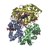
| Details | XC229 was shown to be a homotetramer. However, there was only one subunit per asymmetric unit in the crystals employed in this study. The tetramer was constructed by rotating the contents of the asymmetric unit using the crystallographic I-centered 2-fold symmetry(I23). |
-Components
| #1: Protein | Mass: 15198.329 Da / Num. of mol.: 1 Source method: isolated from a genetically manipulated source Source: (gene. exp.) Xanthomonas campestris pv. campestris (bacteria)Species: Xanthomonas campestris / Strain: ATCC 33913 / Plasmid: pET20b / Species (production host): Escherichia coli / Production host: |
|---|---|
| #2: Water | ChemComp-HOH /  Mass: 18.015 Da / Num. of mol.: 171 / Source method: isolated from a natural source / Formula: H2O Mass: 18.015 Da / Num. of mol.: 171 / Source method: isolated from a natural source / Formula: H2O |
-Experimental details
-Experiment
| Experiment | Method: X-RAY DIFFRACTION / Number of used crystals: 2 |
|---|
- Sample preparation
| Crystal | Density Matthews: 3.32 Å3/Da / Density % sol: 62.94 % |
|---|---|
| Crystal grow | Temperature: 295 K / Method: vapor diffusion, sitting drop / pH: 8 Details: MPD33%, pH 8.0, VAPOR DIFFUSION, SITTING DROP, temperature 295K |
-Data collection
| Diffraction | Mean temperature: 100 K |
|---|---|
| Diffraction source | Source: SYNCHROTRON / Site: SPring-8  / Beamline: BL12B2 / Wavelength: 0.9823 Å / Beamline: BL12B2 / Wavelength: 0.9823 Å |
| Detector | Type: ADSC QUANTUM 4 / Detector: CCD / Date: Jun 3, 2005 |
| Radiation | Monochromator: Se-Met / Protocol: SINGLE WAVELENGTH / Monochromatic (M) / Laue (L): M / Scattering type: x-ray |
| Radiation wavelength | Wavelength: 0.9823 Å / Relative weight: 1 |
| Reflection | Resolution: 1.7→26.6 Å / Num. all: 43207 / Num. obs: 41911 / % possible obs: 97.3 % / Observed criterion σ(F): 2 / Observed criterion σ(I): 2 |
- Processing
| Software |
| ||||||||||||||||||||
|---|---|---|---|---|---|---|---|---|---|---|---|---|---|---|---|---|---|---|---|---|---|
| Refinement | Method to determine structure: SAD / Resolution: 1.7→26.6 Å / σ(F): 2 / Stereochemistry target values: Engh & Huber
| ||||||||||||||||||||
| Refinement step | Cycle: LAST / Resolution: 1.7→26.6 Å
|
