 Movie
Movie Controller


+ Open data

- Basic information
| Entry | Database: PDB / ID: 5m87 | ||||||
|---|---|---|---|---|---|---|---|
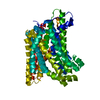
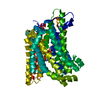
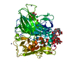
| Title | Crystal structure of Eremococcus coleocola manganese transporter | ||||||
 Components Components | Divalent metal cation transporter MntH | ||||||
 Keywords Keywords | TRANSPORT PROTEIN | ||||||
| Function / homology | NRAMP family / Natural resistance-associated macrophage protein-like / cadmium ion transmembrane transporter activity / manganese ion transmembrane transporter activity / iron ion transmembrane transport / symporter activity / metal ion binding / plasma membrane / Divalent metal cation transporter MntH Function and homology information Function and homology information | ||||||
| Biological species |  Eremococcus coleocola ACS-139-V-Col8 (bacteria) Eremococcus coleocola ACS-139-V-Col8 (bacteria) | ||||||
| Method |  X-RAY DIFFRACTION / SYNCHROTRON / MAD / Resolution: 3.3 Å X-RAY DIFFRACTION / SYNCHROTRON / MAD / Resolution: 3.3 Å | ||||||
 Authors Authors | Manatschal, C. / Ehrnstorfer, I.A. / Arnold, F.M. / Laederach, J. / Dutzler, R. | ||||||
| Funding support |  Switzerland, 1items Switzerland, 1items
| ||||||
 Citation Citation | Journal: Nat Commun / Year: 2017 Title: Structural and mechanistic basis of proton-coupled metal ion transport in the SLC11/NRAMP family. Authors: Ehrnstorfer, I.A. / Manatschal, C. / Arnold, F.M. / Laederach, J. / Dutzler, R. | ||||||
| History |
|
- Structure visualization
| Structure viewer | Molecule: MolmilJmol/JSmol |
|---|
- Downloads & links
-Download
| PDBx/mmCIF format | 5m87.cif.gz | 208.7 KB | Display | PDBx/mmCIF format |
|---|---|---|---|---|
| PDB format | pdb5m87.ent.gz | 169.1 KB | Display | PDB format |
| PDBx/mmJSON format | 5m87.json.gz | Tree view | PDBx/mmJSON format | |
| Others |  Other downloads Other downloads |
-Validation report
| Arichive directory | https://data.pdbj.org/pub/pdb/validation_reports/m8/5m87ftp://data.pdbj.org/pub/pdb/validation_reports/m8/5m87 | HTTPS FTP |
|---|
-Related structure data


-Links
 PDBj
PDBj- Assembly
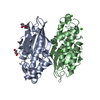
| Deposited unit | 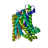
| ||||||||
|---|---|---|---|---|---|---|---|---|---|
| 1 |
| ||||||||
| Unit cell |
|
-Components
| #1: Protein | Mass: 56628.547 Da / Num. of mol.: 1 Source method: isolated from a genetically manipulated source Source: (gene. exp.) Eremococcus coleocola ACS-139-V-Col8 (bacteria)Gene: mntH, HMPREF9257_1603 / Production host: |
|---|---|
| #2: Sugar | ChemComp-DMU /   Type: D-saccharide / Mass: 482.562 Da / Num. of mol.: 1 Type: D-saccharide / Mass: 482.562 Da / Num. of mol.: 1Source method: isolated from a genetically manipulated source Formula: C22H42O11 / Comment: detergent*YM |
-Experimental details
-Experiment
| Experiment | Method: X-RAY DIFFRACTION / Number of used crystals: 1 |
|---|
- Sample preparation
| Crystal | Density Matthews: 5.01 Å3/Da / Density % sol: 75.46 % |
|---|---|
| Crystal grow | Temperature: 277.15 K / Method: vapor diffusion, sitting drop / Details: 50 mM Tris-HCl pH 8.6 22,8% PEG 400 (v/v) |
-Data collection
| Diffraction | Mean temperature: 80 K | |||||||||||||||
|---|---|---|---|---|---|---|---|---|---|---|---|---|---|---|---|---|
| Diffraction source | Source: SYNCHROTRON / Site: SLS / Beamline: X06DA / Wavelength: 1.0, 0.9794, 0.9796, 0.9173 | |||||||||||||||
| Detector | Type: DECTRIS PILATUS 2M-F / Detector: PIXEL / Date: Feb 22, 2016 | |||||||||||||||
| Radiation | Protocol: MAD / Monochromatic (M) / Laue (L): M / Scattering type: x-ray | |||||||||||||||
| Radiation wavelength |
| |||||||||||||||
| Reflection | Resolution: 3.3→50 Å / Num. obs: 16757 / % possible obs: 99.7 % / Redundancy: 13.6 % / CC1/2: 0.998 / Rrim(I) all: 0.051 / Net I/σ(I): 26.6 | |||||||||||||||
| Reflection shell | Resolution: 3.3→3.4 Å / Mean I/σ(I) obs: 1.8 / Num. unique all: 1441 / CC1/2: 0.865 / Rrim(I) all: 1.646 / % possible all: 99.6 |
- Processing
| Software |
| ||||||||||||||||||||||||||||||||||||||||||||||||||||||||||||||||||||||||||||||||||||||||||||||||||||||||||||||||||||||||||||||||||||||||||||||||||||||||||||||||||||||||||||||||||||||||||||||||||||||||||||||||||||||||||||||||||||||||||||||||||||||||||
|---|---|---|---|---|---|---|---|---|---|---|---|---|---|---|---|---|---|---|---|---|---|---|---|---|---|---|---|---|---|---|---|---|---|---|---|---|---|---|---|---|---|---|---|---|---|---|---|---|---|---|---|---|---|---|---|---|---|---|---|---|---|---|---|---|---|---|---|---|---|---|---|---|---|---|---|---|---|---|---|---|---|---|---|---|---|---|---|---|---|---|---|---|---|---|---|---|---|---|---|---|---|---|---|---|---|---|---|---|---|---|---|---|---|---|---|---|---|---|---|---|---|---|---|---|---|---|---|---|---|---|---|---|---|---|---|---|---|---|---|---|---|---|---|---|---|---|---|---|---|---|---|---|---|---|---|---|---|---|---|---|---|---|---|---|---|---|---|---|---|---|---|---|---|---|---|---|---|---|---|---|---|---|---|---|---|---|---|---|---|---|---|---|---|---|---|---|---|---|---|---|---|---|---|---|---|---|---|---|---|---|---|---|---|---|---|---|---|---|---|---|---|---|---|---|---|---|---|---|---|---|---|---|---|---|---|---|---|---|---|---|---|---|---|---|---|---|---|---|---|---|---|
| Refinement | Method to determine structure: MAD / Resolution: 3.3→12 Å / SU ML: 0.41 / Cross valid method: FREE R-VALUE / σ(F): 1.34 / Phase error: 38.61
| ||||||||||||||||||||||||||||||||||||||||||||||||||||||||||||||||||||||||||||||||||||||||||||||||||||||||||||||||||||||||||||||||||||||||||||||||||||||||||||||||||||||||||||||||||||||||||||||||||||||||||||||||||||||||||||||||||||||||||||||||||||||||||
| Solvent computation | Shrinkage radii: 0.9 Å / VDW probe radii: 1.11 Å | ||||||||||||||||||||||||||||||||||||||||||||||||||||||||||||||||||||||||||||||||||||||||||||||||||||||||||||||||||||||||||||||||||||||||||||||||||||||||||||||||||||||||||||||||||||||||||||||||||||||||||||||||||||||||||||||||||||||||||||||||||||||||||
| Refinement step | Cycle: LAST / Resolution: 3.3→12 Å
| ||||||||||||||||||||||||||||||||||||||||||||||||||||||||||||||||||||||||||||||||||||||||||||||||||||||||||||||||||||||||||||||||||||||||||||||||||||||||||||||||||||||||||||||||||||||||||||||||||||||||||||||||||||||||||||||||||||||||||||||||||||||||||
| Refine LS restraints |
| ||||||||||||||||||||||||||||||||||||||||||||||||||||||||||||||||||||||||||||||||||||||||||||||||||||||||||||||||||||||||||||||||||||||||||||||||||||||||||||||||||||||||||||||||||||||||||||||||||||||||||||||||||||||||||||||||||||||||||||||||||||||||||
| LS refinement shell |
| ||||||||||||||||||||||||||||||||||||||||||||||||||||||||||||||||||||||||||||||||||||||||||||||||||||||||||||||||||||||||||||||||||||||||||||||||||||||||||||||||||||||||||||||||||||||||||||||||||||||||||||||||||||||||||||||||||||||||||||||||||||||||||
| Refinement TLS params. | Method: refined / Refine-ID: X-RAY DIFFRACTION
| ||||||||||||||||||||||||||||||||||||||||||||||||||||||||||||||||||||||||||||||||||||||||||||||||||||||||||||||||||||||||||||||||||||||||||||||||||||||||||||||||||||||||||||||||||||||||||||||||||||||||||||||||||||||||||||||||||||||||||||||||||||||||||
| Refinement TLS group |
|
