- PDB-2b0o: Crystal structure of UPLC1 GAP domain -
+
Open data
ID or keywords:
Loading...
-
Basic information
Entry
Database: PDB / ID: 2b0o
Title
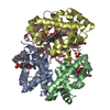
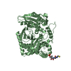
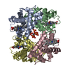
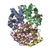
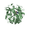
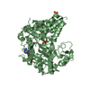
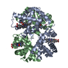
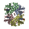
Crystal structure of UPLC1 GAP domain
Components
UPLC1
Keywords
METAL BINDING PROTEIN / ARFGAP / Structural Genomics / Structural Genomics Consortium / SGC
Function / homology
Function and homology information
regulation of stress fiber assembly / positive regulation of GTPase activity / ruffle / GTPase activator activity / cell migration / focal adhesion / zinc ion binding / nucleoplasm / plasma membrane / cytoplasm / cytosol Similarity search - Function
Arf GTPase activating protein, ASAP3 / : / ASAP, PH domain / Arf-GAP with SH3 domain, ANK repeat and PH domain-containing protein / Arf GTPase activating protein / BAR domain of APPL family / Arf GTPase activating protein / ArfGAP domain superfamily / Putative GTPase activating protein for Arf / ARF GTPase-activating proteins domain profile. ...Arf GTPase activating protein, ASAP3 / : / ASAP, PH domain / Arf-GAP with SH3 domain, ANK repeat and PH domain-containing protein / Arf GTPase activating protein / BAR domain of APPL family / Arf GTPase activating protein / ArfGAP domain superfamily / Putative GTPase activating protein for Arf / ARF GTPase-activating proteins domain profile. / Putative GTP-ase activating proteins for the small GTPase, ARF / ARFGAP/RecO-like zinc finger / BAR domain / Annexin V; domain 1 / AH/BAR domain superfamily / Ankyrin repeat-containing domain / PH domain / PH domain profile. / Pleckstrin homology domain. / Pleckstrin homology domain / Ankyrin repeat profile. / Ankyrin repeats (3 copies) / Ankyrin repeat region circular profile. / ankyrin repeats / Ankyrin repeat / Ankyrin repeat-containing domain superfamily / Serine Threonine Protein Phosphatase 5, Tetratricopeptide repeat / Alpha Horseshoe / PH-like domain superfamily / Orthogonal Bundle / Mainly Alpha Similarity search - Domain/homology
Arf-GAP with SH3 domain, ANK repeat and PH domain-containing protein 3 / Arf-GAP with SH3 domain, ANK repeat and PH domain-containing protein 3 Similarity search - Component
In the structure databanks used in Yorodumi, some data are registered as the other names, "COVID-19 virus" and "2019-nCoV". Here are the details of the virus and the list of structure data.
Jan 31, 2019. EMDB accession codes are about to change! (news from PDBe EMDB page)
EMDB accession codes are about to change! (news from PDBe EMDB page)
The allocation of 4 digits for EMDB accession codes will soon come to an end. Whilst these codes will remain in use, new EMDB accession codes will include an additional digit and will expand incrementally as the available range of codes is exhausted. The current 4-digit format prefixed with “EMD-” (i.e. EMD-XXXX) will advance to a 5-digit format (i.e. EMD-XXXXX), and so on. It is currently estimated that the 4-digit codes will be depleted around Spring 2019, at which point the 5-digit format will come into force.
The EM Navigator/Yorodumi systems omit the EMD- prefix.
Related info.:Q: What is EMD? / ID/Accession-code notation in Yorodumi/EM Navigator
Yorodumi is a browser for structure data from EMDB, PDB, SASBDB, etc.
This page is also the successor to EM Navigator detail page, and also detail information page/front-end page for Omokage search.
The word "yorodu" (or yorozu) is an old Japanese word meaning "ten thousand". "mi" (miru) is to see.
Related info.:EMDB / PDB / SASBDB / Comparison of 3 databanks / Yorodumi Search / Aug 31, 2016. New EM Navigator & Yorodumi / Yorodumi Papers / Jmol/JSmol / Function and homology information / Changes in new EM Navigator and Yorodumi
 Movie
Movie Controller


 Open data
 Basic information
 Components
 Keywords
 Function and homology information
 Homo sapiens (human)
 X-RAY DIFFRACTION /
  Authors
 Citation
 Structure visualization
 Downloads & links
 Other downloads
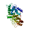


 PDBj
PDBj

 Assembly
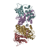

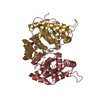


 Mass: 65.409 Da / Num. of mol.: 4 / Source method: obtained synthetically / Formula: Zn
 Mass: 18.015 Da / Num. of mol.: 500 / Source method: isolated from a natural source / Formula: H2O
 Sample preparation
 / Beamline: X29A / Wavelength: 0.979 Å
 Processing