Movie
Movie Controller

[English] 日本語
 Yorodumi
- PDB-2ftc: Structural Model for the Large Subunit of the Mammalian Mitochond... -
+ Open data

- Basic information
| Entry | Database: PDB / ID: 2ftc | ||||||
|---|---|---|---|---|---|---|---|
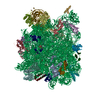
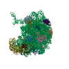
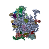
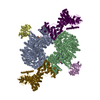
| Title | Structural Model for the Large Subunit of the Mammalian Mitochondrial Ribosome | ||||||
 Components Components |
| ||||||
 Keywords Keywords | RIBOSOME / MITOCHONDRIAL RIBOSOME / LARGE RIBOSOMAL SUBUNIT / RIBOSOMAL RNA | ||||||
| Function / homology |  Function and homology information Function and homology informationMitochondrial translation elongation / Mitochondrial ribosome-associated quality control / mitochondrial transcription / mitochondrial large ribosomal subunit / Mitochondrial translation termination / mitochondrial ribosome / mitochondrial translation / Mitochondrial protein degradation / large ribosomal subunit / large ribosomal subunit rRNA binding ...Mitochondrial translation elongation / Mitochondrial ribosome-associated quality control / mitochondrial transcription / mitochondrial large ribosomal subunit / Mitochondrial translation termination / mitochondrial ribosome / mitochondrial translation / Mitochondrial protein degradation / large ribosomal subunit / large ribosomal subunit rRNA binding / mitochondrial inner membrane / negative regulation of translation / rRNA binding / structural constituent of ribosome / ribosome / translation / mitochondrial matrix / mRNA binding / positive regulation of DNA-templated transcription / mitochondrion / RNA binding Similarity search - Function | ||||||
| Biological species |  | ||||||
| Method | ELECTRON MICROSCOPY / single particle reconstruction / cryo EM / Resolution: 12.1 Å | ||||||
 Authors Authors | Mears, J.A. / Sharma, M.R. / Gutell, R.R. / Richardson, P.E. / Agrawal, R.K. / Harvey, S.C. | ||||||
 Citation Citation | Journal: J Mol Biol / Year: 2006 Title: A structural model for the large subunit of the mammalian mitochondrial ribosome. Authors: Jason A Mears / Manjuli R Sharma / Robin R Gutell / Amanda S McCook / Paul E Richardson / Thomas R Caulfield / Rajendra K Agrawal / Stephen C Harvey /  Abstract: Protein translation is essential for all forms of life and is conducted by a macromolecular complex, the ribosome. Evolutionary changes in protein and RNA sequences can affect the 3D organization of ...Protein translation is essential for all forms of life and is conducted by a macromolecular complex, the ribosome. Evolutionary changes in protein and RNA sequences can affect the 3D organization of structural features in ribosomes in different species. The most dramatic changes occur in animal mitochondria, whose genomes have been reduced and altered significantly. The RNA component of the mitochondrial ribosome (mitoribosome) is reduced in size, with a compensatory increase in protein content. Until recently, it was unclear how these changes affect the 3D structure of the mitoribosome. Here, we present a structural model of the large subunit of the mammalian mitoribosome developed by combining molecular modeling techniques with cryo-electron microscopic data at 12.1A resolution. The model contains 93% of the mitochondrial rRNA sequence and 16 mitochondrial ribosomal proteins in the large subunit of the mitoribosome. Despite the smaller mitochondrial rRNA, the spatial positions of RNA domains known to be involved directly in protein synthesis are essentially the same as in bacterial and archaeal ribosomes. However, the dramatic reduction in rRNA content necessitates evolution of unique structural features to maintain connectivity between RNA domains. The smaller rRNA sequence also limits the likelihood of tRNA binding at the E-site of the mitoribosome, and correlates with the reduced size of D-loops and T-loops in some animal mitochondrial tRNAs, suggesting co-evolution of mitochondrial rRNA and tRNA structures. | ||||||
| History |
| ||||||
| Remark 999 | SEQUENCE SINCE THE SEQUENCE OF THE HUMAN AND BOVINE MITOCHONDRIAL PROTEINS ARE ESSENTIALLY ...SEQUENCE SINCE THE SEQUENCE OF THE HUMAN AND BOVINE MITOCHONDRIAL PROTEINS ARE ESSENTIALLY IDENTICAL, THE SEQUENCE DATABASE REFERENCE FOR THE PROTEINS USED IN MODELLING FOR THIS DEPOSITION WAS FROM A HUNAN SOURCE (AND NOT BOVINE).THUS THE REFERENCE USED FOR THE PROTEIN CHAINS ARE LISTED BELOW CHAIN ID DATABASE REFERENCE A GB AAH14356 B GB NP_057034 C SWS RM03_HUMAN D GB NP_666499 E GB NP_963900 F GB NP_963900 G SWS RM11_HUMAN H SWS RM13_HUMAN I GB AAH01040 J GB AAH12306 K SWS RM19_HUMAN L GB AAH59945 M GB NP_054899 N GB CAI16343 O SWS RM27_HUMAN P GB NP_004882 Q SWS RM34_HUMAN |
- Structure visualization
| Movie |
Movie viewer |
|---|---|
| Structure viewer | Molecule: MolmilJmol/JSmol |
- Downloads & links
-Download
| PDBx/mmCIF format | 2ftc.cif.gz | 133.6 KB | Display | PDBx/mmCIF format |
|---|---|---|---|---|
| PDB format | pdb2ftc.ent.gz | 71.5 KB | Display | PDB format |
| PDBx/mmJSON format | 2ftc.json.gz | Tree view | PDBx/mmJSON format | |
| Others |  Other downloads Other downloads |
-Validation report
| Arichive directory | https://data.pdbj.org/pub/pdb/validation_reports/ft/2ftcftp://data.pdbj.org/pub/pdb/validation_reports/ft/2ftc | HTTPS FTP |
|---|
-Related structure data
| Similar structure data |
|---|


-Links
 PDBj
PDBj


- Assembly
| Deposited unit | 
|
|---|---|
| 1 |
|
-Components
-Mitochondrial ribosomal protein ... , 8 types, 8 molecules ABDIJMNP
| #2: Protein | Mass: 21224.551 Da / Num. of mol.: 1 / Source method: isolated from a natural source / Source: (natural) |
|---|---|
| #3: Protein | Mass: 14442.636 Da / Num. of mol.: 1 / Source method: isolated from a natural source / Source: (natural) |
| #5: Protein | Mass: 19707.887 Da / Num. of mol.: 1 / Source method: isolated from a natural source / Source: (natural) |
| #9: Protein | Mass: 13107.179 Da / Num. of mol.: 1 / Source method: isolated from a natural source / Source: (natural) |
| #10: Protein | Mass: 13682.971 Da / Num. of mol.: 1 / Source method: isolated from a natural source / Source: (natural) |
| #13: Protein | Mass: 12914.133 Da / Num. of mol.: 1 / Source method: isolated from a natural source / Source: (natural) |
| #14: Protein | Mass: 11010.680 Da / Num. of mol.: 1 / Source method: isolated from a natural source / Source: (natural) |
| #16: Protein | Mass: 6092.276 Da / Num. of mol.: 1 / Source method: isolated from a natural source / Source: (natural) |
-Mitochondrial 39S ribosomal protein ... , 2 types, 2 molecules CO
| #4: Protein | Mass: 23396.496 Da / Num. of mol.: 1 / Source method: isolated from a natural source / Source: (natural) |
|---|---|
| #15: Protein | Mass: 7520.631 Da / Num. of mol.: 1 / Source method: isolated from a natural source / Source: (natural) |
-39S ribosomal protein ... , 5 types, 6 molecules EFGHKQ
| #6: Protein | Mass: 14831.392 Da / Num. of mol.: 2 / Source method: isolated from a natural source / Source: (natural) #7: Protein | | Mass: 15606.364 Da / Num. of mol.: 1 / Source method: isolated from a natural source / Source: (natural) #8: Protein | | Mass: 16958.729 Da / Num. of mol.: 1 / Source method: isolated from a natural source / Source: (natural) #11: Protein | | Mass: 11098.766 Da / Num. of mol.: 1 / Source method: isolated from a natural source / Source: (natural) #17: Protein/peptide | | Mass: 4485.404 Da / Num. of mol.: 1 / Source method: isolated from a natural source / Source: (natural) |
|---|
-RNA chain / Protein , 2 types, 2 molecules RL
| #12: Protein | Mass: 14125.866 Da / Num. of mol.: 1 / Source method: isolated from a natural source / Source: (natural) |
|---|---|
| #1: RNA chain | Mass: 504151.281 Da / Num. of mol.: 1 / Source method: isolated from a natural source / Source: (natural) |
-Experimental details
-Experiment
| Experiment | Method: ELECTRON MICROSCOPY |
|---|---|
| EM experiment | Aggregation state: PARTICLE / 3D reconstruction method: single particle reconstruction |
- Sample preparation
| Component | Name: Mammalian Mitochondrial Ribosome Large Subunit / Type: RIBOSOME |
|---|---|
| Specimen | Embedding applied: NO / Shadowing applied: NO / Staining applied: NO / Vitrification applied: YES |
- Electron microscopy imaging
| Experimental equipment |  Model: Tecnai F20 / Image courtesy: FEI Company |
|---|---|
| Microscopy | Model: FEI TECNAI F20 |
| Electron gun | Electron source:  FIELD EMISSION GUN / Accelerating voltage: 200 kV / Illumination mode: FLOOD BEAM FIELD EMISSION GUN / Accelerating voltage: 200 kV / Illumination mode: FLOOD BEAM |
| Electron lens | Mode: BRIGHT FIELD / Nominal defocus max: 4600 nm / Nominal defocus min: 1200 nm |
| Image scans | Num. digital images: 194 / Scanner model: ZEISS SCAI |
- Processing
| Software | Name: YAMMP / Classification: refinement | |||||||||||||||||||||||||||||||||||
|---|---|---|---|---|---|---|---|---|---|---|---|---|---|---|---|---|---|---|---|---|---|---|---|---|---|---|---|---|---|---|---|---|---|---|---|---|
| Symmetry | Point symmetry: C1 (asymmetric) | |||||||||||||||||||||||||||||||||||
| 3D reconstruction | Resolution: 12.1 Å / Symmetry type: POINT | |||||||||||||||||||||||||||||||||||
| Atomic model building | Protocol: RIGID BODY FIT / Space: REAL / Details: REFINEMENT PROTOCOL--rigid body | |||||||||||||||||||||||||||||||||||
| Atomic model building |
| |||||||||||||||||||||||||||||||||||
| Refinement step | Cycle: LAST
|