 Movie
Movie Controller


+ Open data

- Basic information
| Entry | Database: PDB / ID: 2frv | ||||||
|---|---|---|---|---|---|---|---|
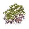
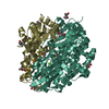
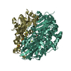
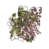
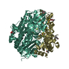
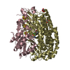
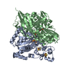
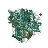
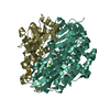
| Title | CRYSTAL STRUCTURE OF THE OXIDIZED FORM OF NI-FE HYDROGENASE | ||||||
 Components Components | (PERIPLASMIC HYDROGENASE) x 2 | ||||||
 Keywords Keywords | OXIDOREDUCTASE / NI-FE HYDROGENASE | ||||||
| Function / homology |  Function and homology information Function and homology informationcytochrome-c3 hydrogenase / cytochrome-c3 hydrogenase activity / ferredoxin hydrogenase complex / [Ni-Fe] hydrogenase complex / ferredoxin hydrogenase activity / anaerobic respiration / 3 iron, 4 sulfur cluster binding / nickel cation binding / 4 iron, 4 sulfur cluster binding / electron transfer activity ...cytochrome-c3 hydrogenase / cytochrome-c3 hydrogenase activity / ferredoxin hydrogenase complex / [Ni-Fe] hydrogenase complex / ferredoxin hydrogenase activity / anaerobic respiration / 3 iron, 4 sulfur cluster binding / nickel cation binding / 4 iron, 4 sulfur cluster binding / electron transfer activity / periplasmic space / membrane / metal ion binding Similarity search - Function | ||||||
| Biological species |  Desulfovibrio gigas (bacteria) Desulfovibrio gigas (bacteria) | ||||||
| Method |  X-RAY DIFFRACTION / SYNCHROTRON / MOLECULAR REPLACEMENT / Resolution: 2.54 Å X-RAY DIFFRACTION / SYNCHROTRON / MOLECULAR REPLACEMENT / Resolution: 2.54 Å | ||||||
 Authors Authors | Volbeda, A. / Frey, M. / Fontecilla-Camps, J.C. | ||||||
 Citation Citation | Journal: J.Am.Chem.Soc. / Year: 1996 Title: Structure of the [Nife] Hydrogenase Active Site: Evidence for Biologically Uncommon Fe Ligands Authors: Volbeda, A. / Garcin, E. / Piras, C. / De Lacey, A.L. / Fernandez, V.M. / Hatchikian, E.C. / Frey, M. / Fontecilla-Camps, J.C. #1: Journal: Nat.Struct.Biol. / Year: 1997Title: Gas Access to the Active Site of Ni-Fe Hydrogenases Probed by X-Ray Crystallography and Molecular Dynamics Authors: Montet, Y. / Amara, P. / Volbeda, A. / Vernede, X. / Hatchikian, E.C. / Field, M.J. / Frey, M. / Fontecilla-Camps, J.C. #2: Journal: Nature / Year: 1995Title: Crystal Structure of the Nickel-Iron Hydrogenase from Desulfovibrio Gigas Authors: Volbeda, A. / Charon, M.H. / Piras, C. / Hatchikian, E.C. / Frey, M. / Fontecilla-Camps, J.C. | ||||||
| History |
|
- Structure visualization
| Structure viewer | Molecule: MolmilJmol/JSmol |
|---|
- Downloads & links
-Download
| PDBx/mmCIF format | 2frv.cif.gz | 909.7 KB | Display | PDBx/mmCIF format |
|---|---|---|---|---|
| PDB format | pdb2frv.ent.gz | 741.6 KB | Display | PDB format |
| PDBx/mmJSON format | 2frv.json.gz | Tree view | PDBx/mmJSON format | |
| Others |  Other downloads Other downloads |
-Validation report
| Arichive directory | https://data.pdbj.org/pub/pdb/validation_reports/fr/2frvftp://data.pdbj.org/pub/pdb/validation_reports/fr/2frv | HTTPS FTP |
|---|
-Related structure data
| Related structure data |  1frvS S: Starting model for refinement |
|---|---|
| Similar structure data |

-Links
 PDBj
PDBj


- Assembly
| Deposited unit | 
| ||||||||||||||||||||||||
|---|---|---|---|---|---|---|---|---|---|---|---|---|---|---|---|---|---|---|---|---|---|---|---|---|---|
| 1 | 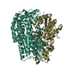
| ||||||||||||||||||||||||
| 2 | 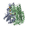
| ||||||||||||||||||||||||
| 3 | 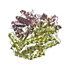
| ||||||||||||||||||||||||
| 4 | 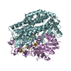
| ||||||||||||||||||||||||
| 5 | 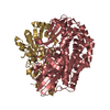
| ||||||||||||||||||||||||
| 6 | 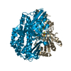
| ||||||||||||||||||||||||
| Unit cell |
| ||||||||||||||||||||||||
| Noncrystallographic symmetry (NCS) | NCS oper:
| ||||||||||||||||||||||||
| Details | THERE ARE SIX MOLECULES PER (TRICLINIC) UNIT CELL. EACH OF THESE CONSISTS OF ONE SMALL AND ONE LARGE SUBUNIT. IN ALL STAGES THE SIX HYDROGENASE MOLECULES WERE CONSTRAINED TO OBEY EXACT NON-CRYSTALLOGRAPHIC SYMMETRY. |
-Components
-Protein , 2 types, 12 molecules SACEGILBDFHJ
| #1: Protein | Mass: 28351.396 Da / Num. of mol.: 6 / Source method: isolated from a natural source / Source: (natural) Desulfovibrio gigas (bacteria) / Cellular location: PERIPLASM / References: UniProt: P12943, 1.18.99.1#2: Protein | Mass: 59571.938 Da / Num. of mol.: 6 / Source method: isolated from a natural source / Source: (natural) Desulfovibrio gigas (bacteria) / Cellular location: PERIPLASM / References: UniProt: P12944, 1.18.99.1 |
|---|
-Non-polymers , 7 types, 1188 molecules 


| #3: Chemical | ChemComp-SF4 /  Mass: 351.640 Da / Num. of mol.: 12 / Source method: obtained synthetically / Formula: Fe4S4 Mass: 351.640 Da / Num. of mol.: 12 / Source method: obtained synthetically / Formula: Fe4S4#4: Chemical | ChemComp-F3S /  Mass: 295.795 Da / Num. of mol.: 6 / Source method: obtained synthetically / Formula: Fe3S4 Mass: 295.795 Da / Num. of mol.: 6 / Source method: obtained synthetically / Formula: Fe3S4#5: Chemical | ChemComp-NI /  Mass: 58.693 Da / Num. of mol.: 6 / Source method: obtained synthetically / Formula: Ni Mass: 58.693 Da / Num. of mol.: 6 / Source method: obtained synthetically / Formula: Ni#6: Chemical | ChemComp-MG /  Mass: 24.305 Da / Num. of mol.: 6 / Source method: obtained synthetically / Formula: Mg Mass: 24.305 Da / Num. of mol.: 6 / Source method: obtained synthetically / Formula: Mg#7: Chemical | ChemComp-FCO /  Mass: 135.890 Da / Num. of mol.: 6 / Source method: obtained synthetically / Formula: C3FeN2O Mass: 135.890 Da / Num. of mol.: 6 / Source method: obtained synthetically / Formula: C3FeN2O#8: Chemical | ChemComp-O /  Mass: 15.999 Da / Num. of mol.: 6 / Source method: obtained synthetically / Formula: O Mass: 15.999 Da / Num. of mol.: 6 / Source method: obtained synthetically / Formula: O#9: Water | ChemComp-HOH / | Mass: 18.015 Da / Num. of mol.: 1146 / Source method: isolated from a natural source / Formula: H2O |
|---|
-Details
| Has protein modification | Y |
|---|---|
| Nonpolymer details | THE ACTIVE SITE CONTAINS TWO METALS: NI AND FE. THE LATTER ASSIGNMENT HAS BEEN CONFIRMED BY ...THE ACTIVE SITE CONTAINS TWO METALS: NI AND FE. THE LATTER ASSIGNMENT |
-Experimental details
-Experiment
| Experiment | Method: X-RAY DIFFRACTION / Number of used crystals: 1 |
|---|
- Sample preparation
| Crystal | Density Matthews: 2.8 Å3/Da / Density % sol: 56.14 % |
|---|---|
| Crystal grow | pH: 6.6 / Details: pH 6.6 |
| Crystal grow | *PLUS Method: unknown |
-Data collection
| Diffraction | Mean temperature: 120 K |
|---|---|
| Diffraction source | Source: SYNCHROTRON / Site: ESRF  / Beamline: ID2 / Wavelength: 0.905 / Beamline: ID2 / Wavelength: 0.905 |
| Detector | Type: MARRESEARCH / Detector: IMAGE PLATE / Date: Jun 1, 1994 |
| Radiation | Monochromatic (M) / Laue (L): M / Scattering type: x-ray |
| Radiation wavelength | Wavelength: 0.905 Å / Relative weight: 1 |
| Reflection | Resolution: 2.54→29.36 Å / Num. obs: 174383 / % possible obs: 92.5 % / Observed criterion σ(I): 4 / Redundancy: 1.7 % / Biso Wilson estimate: 7.3 Å2 / Rmerge(I) obs: 0.066 / Net I/σ(I): 7.5 |
| Reflection shell | Resolution: 2.54→2.84 Å / Redundancy: 1.57 % / Rmerge(I) obs: 0.151 / Mean I/σ(I) obs: 4 / % possible all: 87.4 |
| Reflection | *PLUS Num. measured all: 296283 |
| Reflection shell | *PLUS % possible obs: 87.4 % |
- Processing
| Software |
| ||||||||||||||||||||||||||||||||||||||||||||||||||||||||||||||||||||||||||||||||||||
|---|---|---|---|---|---|---|---|---|---|---|---|---|---|---|---|---|---|---|---|---|---|---|---|---|---|---|---|---|---|---|---|---|---|---|---|---|---|---|---|---|---|---|---|---|---|---|---|---|---|---|---|---|---|---|---|---|---|---|---|---|---|---|---|---|---|---|---|---|---|---|---|---|---|---|---|---|---|---|---|---|---|---|---|---|---|
| Refinement | Method to determine structure: MOLECULAR REPLACEMENT Starting model: PDB ENTRY 1FRV, MOLECULE 1 Resolution: 2.54→8 Å / Cross valid method: R-FREE / σ(F): 0 / Details: BEFORE PROLSQ: AMORE + X-PLOR
| ||||||||||||||||||||||||||||||||||||||||||||||||||||||||||||||||||||||||||||||||||||
| Displacement parameters | Biso mean: 9.2 Å2 | ||||||||||||||||||||||||||||||||||||||||||||||||||||||||||||||||||||||||||||||||||||
| Refine analyze | Luzzati d res low obs: 20 Å / Luzzati sigma a obs: 0.27 Å | ||||||||||||||||||||||||||||||||||||||||||||||||||||||||||||||||||||||||||||||||||||
| Refinement step | Cycle: LAST / Resolution: 2.54→8 Å
| ||||||||||||||||||||||||||||||||||||||||||||||||||||||||||||||||||||||||||||||||||||
| Refine LS restraints |
|
