 Movie
Movie Controller


+ Open data

- Basic information
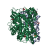
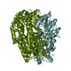
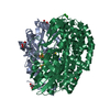
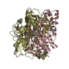
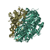
| Entry | Database: PDB / ID: 1frv | ||||||
|---|---|---|---|---|---|---|---|
| Title | CRYSTAL STRUCTURE OF THE OXIDIZED FORM OF NI-FE HYDROGENASE | ||||||
 Components Components | (HYDROGENASE) x 2 | ||||||
 Keywords Keywords | OXIDOREDUCTASE / NI-FE HYDROGENASE | ||||||
| Function / homology |  Function and homology information Function and homology informationcytochrome-c3 hydrogenase / cytochrome-c3 hydrogenase activity / ferredoxin hydrogenase complex / [Ni-Fe] hydrogenase complex / ferredoxin hydrogenase activity / anaerobic respiration / 3 iron, 4 sulfur cluster binding / nickel cation binding / 4 iron, 4 sulfur cluster binding / periplasmic space ...cytochrome-c3 hydrogenase / cytochrome-c3 hydrogenase activity / ferredoxin hydrogenase complex / [Ni-Fe] hydrogenase complex / ferredoxin hydrogenase activity / anaerobic respiration / 3 iron, 4 sulfur cluster binding / nickel cation binding / 4 iron, 4 sulfur cluster binding / periplasmic space / electron transfer activity / membrane / metal ion binding Similarity search - Function | ||||||
| Biological species |  Desulfovibrio gigas (bacteria) Desulfovibrio gigas (bacteria) | ||||||
| Method |  X-RAY DIFFRACTION / SYNCHROTRON / MIR WITH AVERAGING / Resolution: 2.85 Å X-RAY DIFFRACTION / SYNCHROTRON / MIR WITH AVERAGING / Resolution: 2.85 Å | ||||||
 Authors Authors | Volbeda, A. / Frey, M. / Fontecilla-Camps, J.C. | ||||||
 Citation Citation | Journal: Nature / Year: 1995 Title: Crystal structure of the nickel-iron hydrogenase from Desulfovibrio gigas. Authors: Volbeda, A. / Charon, M.H. / Piras, C. / Hatchikian, E.C. / Frey, M. / Fontecilla-Camps, J.C. #1: Journal: Joint Ccp4 Esf-Eacbm Newsletter on Protein CrystallographyYear: 1993 Title: Location of the Redox Centers in Hydrogenase as Determined by X-Ray Crystallography at 5 Angstroms Resolution Authors: Volbeda, A. / Piras, C. / Charon, M.H. / Hatchikian, E.C. / Frey, M. / Fontecilla-Camps, J.C. #2: Journal: J.Bacteriol. / Year: 1989Title: Analysis and Comparison of Nucleotide Sequences Encoding the Genes for [Nife] and [Nifese] Hydrogenases from Desulfovibrio Gigas and Desulfovibrio Baculatus Authors: Voordouw, G. / Menon, N.K. / Legall, J. / Choi, E.S. / Peck Junior, H.D. / Przybyla, A.E. | ||||||
| History |
|
- Structure visualization
| Structure viewer | Molecule: MolmilJmol/JSmol |
|---|
- Downloads & links
-Download
| PDBx/mmCIF format | 1frv.cif.gz | 313.2 KB | Display | PDBx/mmCIF format |
|---|---|---|---|---|
| PDB format | pdb1frv.ent.gz | 250.7 KB | Display | PDB format |
| PDBx/mmJSON format | 1frv.json.gz | Tree view | PDBx/mmJSON format | |
| Others |  Other downloads Other downloads |
-Validation report
| Arichive directory | https://data.pdbj.org/pub/pdb/validation_reports/fr/1frvftp://data.pdbj.org/pub/pdb/validation_reports/fr/1frv | HTTPS FTP |
|---|
-Related structure data
| Similar structure data |
|---|


-Links
 PDBj
PDBj


- Assembly
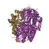
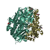
| Deposited unit | 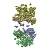
| ||||||||
|---|---|---|---|---|---|---|---|---|---|
| 1 | 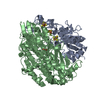
| ||||||||
| 2 | 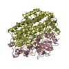
| ||||||||
| Unit cell |
| ||||||||
| Noncrystallographic symmetry (NCS) | NCS oper: (Code: given Matrix: (-0.534696, 0.843915, -0.043675), Vector: |
-Components
-Protein , 2 types, 4 molecules ACBD
| #1: Protein | Mass: 28465.564 Da / Num. of mol.: 2 / Source method: isolated from a natural source / Source: (natural) Desulfovibrio gigas (bacteria) / References: UniProt: P12943, cytochrome-c3 hydrogenase#2: Protein | Mass: 59783.125 Da / Num. of mol.: 2 / Source method: isolated from a natural source / Source: (natural) Desulfovibrio gigas (bacteria) / References: UniProt: P12944, cytochrome-c3 hydrogenase |
|---|
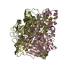
-Non-polymers , 4 types, 10 molecules 


| #3: Chemical | ChemComp-SF4 /  Mass: 351.640 Da / Num. of mol.: 4 / Source method: obtained synthetically / Formula: Fe4S4 Mass: 351.640 Da / Num. of mol.: 4 / Source method: obtained synthetically / Formula: Fe4S4#4: Chemical |  Mass: 295.795 Da / Num. of mol.: 2 / Source method: obtained synthetically / Formula: Fe3S4 Mass: 295.795 Da / Num. of mol.: 2 / Source method: obtained synthetically / Formula: Fe3S4#5: Chemical |  Mass: 58.693 Da / Num. of mol.: 2 / Source method: obtained synthetically / Formula: Ni Mass: 58.693 Da / Num. of mol.: 2 / Source method: obtained synthetically / Formula: Ni#6: Chemical |  Mass: 109.891 Da / Num. of mol.: 2 / Source method: obtained synthetically / Formula: FeH6O3 Mass: 109.891 Da / Num. of mol.: 2 / Source method: obtained synthetically / Formula: FeH6O3 |
|---|
-Details
| Has protein modification | Y |
|---|---|
| Nonpolymer details | THE SMALL SUBUNIT CONTAINS THREE IRON SULFUR CLUSTERS: [4FE-4S](265), [3FE-4S](266) AND [4FE-4S] ...THE SMALL SUBUNIT CONTAINS THREE IRON SULFUR CLUSTERS: [4FE-4S](265), [3FE-4S](266) AND [4FE-4S](267), WITH THE FOLLOWING PROTEIN LIGANDS: CYS 17, CYS 20, CYS 112 AND CYS 148 OF [4FE-4S](269) CYS 228, CYS 46 AND CYS 249 OF [3FE-4S](268) HIS 185, CYS 188, CYS 213 AND CYS 219 OF [4FE-4S](267) |
| Sequence details | THE 15 C-TERMINAL AMINO ACID RESIDUES OF THE LARGE SUBUNIT ARE ABSENT, CONSISTENT WITH ITS REPORTED ...THE 15 C-TERMINAL AMINO ACID RESIDUES OF THE LARGE SUBUNIT ARE ABSENT, CONSISTENT |
-Experimental details
-Experiment
| Experiment | Method: X-RAY DIFFRACTION / Number of used crystals: 1 |
|---|
- Sample preparation
| Crystal | Density Matthews: 2.28 Å3/Da / Density % sol: 46.08 % |
|---|---|
| Crystal grow | pH: 6.6 / Details: pH 6.6 |
| Crystal grow | *PLUS Method: unknown |
-Data collection
| Diffraction | Mean temperature: 293 K |
|---|---|
| Diffraction source | Source: SYNCHROTRON / Site: LURE  / Beamline: DW32 / Wavelength: 0.9 / Beamline: DW32 / Wavelength: 0.9 |
| Detector | Detector: IMAGE PLATE / Date: Sep 23, 1992 |
| Radiation | Monochromatic (M) / Laue (L): M / Scattering type: x-ray |
| Radiation wavelength | Wavelength: 0.9 Å / Relative weight: 1 |
| Reflection | Resolution: 2.85→23.5 Å / Num. obs: 31053 / % possible obs: 85.5 % / Redundancy: 3.7 % / Rmerge(I) obs: 0.086 |
| Reflection shell | Resolution: 2.85→3 Å / Redundancy: 1.6 % / Rmerge(I) obs: 0.207 / % possible all: 47.6 |
| Reflection | *PLUS Num. measured all: 116003 |
- Processing
| Software |
| ||||||||||||||||||||||||||||||||||||||||||||||||||||||||||||||||||||||||||||||||||||
|---|---|---|---|---|---|---|---|---|---|---|---|---|---|---|---|---|---|---|---|---|---|---|---|---|---|---|---|---|---|---|---|---|---|---|---|---|---|---|---|---|---|---|---|---|---|---|---|---|---|---|---|---|---|---|---|---|---|---|---|---|---|---|---|---|---|---|---|---|---|---|---|---|---|---|---|---|---|---|---|---|---|---|---|---|---|
| Refinement | Method to determine structure: MIR WITH AVERAGING / Resolution: 2.85→8 Å / σ(F): 0 Details: ALTHOUGH THE TWO HYDROGENASE MOLECULES WERE REFINED INDEPENDENTLY, DUE TO THE LIMITED RESOLUTION OF THE DATA IT CANNOT BE CONCLUDED THAT, APART FROM LATTICE CONTACT REGIONS, ANY DIFFERENCES ...Details: ALTHOUGH THE TWO HYDROGENASE MOLECULES WERE REFINED INDEPENDENTLY, DUE TO THE LIMITED RESOLUTION OF THE DATA IT CANNOT BE CONCLUDED THAT, APART FROM LATTICE CONTACT REGIONS, ANY DIFFERENCES BETWEEN THE TWO MODELS ARE REAL. THERE IS NO ELECTRON DENSITY FOR RESIDUES 1 - 2 OF THE SMALL SUBUNIT AND RESIDUES 2 - 6 OF THE LARGE SUBUNIT. MET 1 OF THE LARGE SUBUNIT IS ABSENT FROM THE SWISSPROT ENTRY. THE DEPOSITORS KEPT IT SINCE IT WAS INCLUDED IN THE SEQUENCE PUBLICATION (REF. 2), AND DELETING IT WOULD NECESSITATE A RENUMBERING OF ALL RESIDUES. INCIDENTALLY, THE APPARENT ABSENCE OF MET 1 IN THE LARGE SUBUNIT REDUCES THE MENTIONED DISCREPANCY BETWEEN CALCULATED AND OBSERVED MOLECULAR WEIGHT (FOUND BY MASS SPECTROMETRY, SEE NATURE REFERENCE) FROM 245 TO 114 DALTON, WHICH, HOWEVER, STILL IMPLIES A FEW MINOR SEQUENCE ERRORS.
| ||||||||||||||||||||||||||||||||||||||||||||||||||||||||||||||||||||||||||||||||||||
| Displacement parameters | Biso mean: 19 Å2 | ||||||||||||||||||||||||||||||||||||||||||||||||||||||||||||||||||||||||||||||||||||
| Refine analyze | Luzzati coordinate error obs: 0.3 Å | ||||||||||||||||||||||||||||||||||||||||||||||||||||||||||||||||||||||||||||||||||||
| Refinement step | Cycle: LAST / Resolution: 2.85→8 Å
| ||||||||||||||||||||||||||||||||||||||||||||||||||||||||||||||||||||||||||||||||||||
| Refine LS restraints |
| ||||||||||||||||||||||||||||||||||||||||||||||||||||||||||||||||||||||||||||||||||||
| Software | *PLUS Name: PROLSQ / Classification: refinement | ||||||||||||||||||||||||||||||||||||||||||||||||||||||||||||||||||||||||||||||||||||
| Refinement | *PLUS Rfactor obs: 0.178 | ||||||||||||||||||||||||||||||||||||||||||||||||||||||||||||||||||||||||||||||||||||
| Solvent computation | *PLUS | ||||||||||||||||||||||||||||||||||||||||||||||||||||||||||||||||||||||||||||||||||||
| Displacement parameters | *PLUS |
