 Movie
Movie Controller

[English] 日本語
 Yorodumi
- PDB-2fn1: Crystal structures of Yersinia enterocolitica salicylate synthase... -
+ Open data

- Basic information
| Entry | Database: PDB / ID: 2fn1 | |||||||||
|---|---|---|---|---|---|---|---|---|---|---|
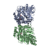
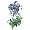
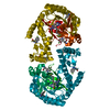
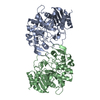
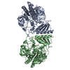
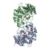
| Title | Crystal structures of Yersinia enterocolitica salicylate synthase (Irp9) in complex with the reaction products salicylate and pyruvate | |||||||||
 Components Components | salicylate synthetase, Irp9 | |||||||||
 Keywords Keywords | TRANSCRIPTION / Yersinia enterocolitica / Irp9 / salicylate synthase / salicylate | |||||||||
| Function / homology |  Function and homology information Function and homology informationcarbon-oxygen lyase activity / oxo-acid-lyase activity / isochorismate synthase activity / siderophore biosynthetic process / L-tryptophan biosynthetic process / magnesium ion binding Similarity search - Function | |||||||||
| Biological species |  Yersinia enterocolitica (bacteria) Yersinia enterocolitica (bacteria) | |||||||||
| Method |  X-RAY DIFFRACTION / MOLECULAR REPLACEMENT / Resolution: 2.1 Å X-RAY DIFFRACTION / MOLECULAR REPLACEMENT / Resolution: 2.1 Å | |||||||||
 Authors Authors | Kerbarh, O. / Chirgadze, D.Y. / Blundell, T.L. / Abell, C. | |||||||||
 Citation Citation | Journal: J.Mol.Biol. / Year: 2006 Title: Crystal Structures of Yersinia enterocolitica Salicylate Synthase and its Complex with the Reaction Products Salicylate and Pyruvate. Authors: Kerbarh, O. / Chirgadze, D.Y. / Blundell, T.L. / Abell, C. | |||||||||
| History |
|
- Structure visualization
| Structure viewer | Molecule: MolmilJmol/JSmol |
|---|
- Downloads & links
-Download
| PDBx/mmCIF format | 2fn1.cif.gz | 177.9 KB | Display | PDBx/mmCIF format |
|---|---|---|---|---|
| PDB format | pdb2fn1.ent.gz | 136.6 KB | Display | PDB format |
| PDBx/mmJSON format | 2fn1.json.gz | Tree view | PDBx/mmJSON format | |
| Others |  Other downloads Other downloads |
-Validation report
| Arichive directory | https://data.pdbj.org/pub/pdb/validation_reports/fn/2fn1ftp://data.pdbj.org/pub/pdb/validation_reports/fn/2fn1 | HTTPS FTP |
|---|
-Related structure data
| Related structure data | 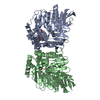 2fn0C 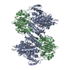 1i1qS  1i7qS 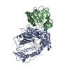 1qdlS C: citing same article ( S: Starting model for refinement |
|---|---|
| Similar structure data |


-Links
 PDBj
PDBj


- Assembly
| Deposited unit | 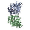
| ||||||||
|---|---|---|---|---|---|---|---|---|---|
| 1 |
| ||||||||
| Unit cell |
| ||||||||
| Details | The biological assembly is the dimer present in the asymmetric unit |
-Components
| #1: Protein | Mass: 48308.910 Da / Num. of mol.: 2 Source method: isolated from a genetically manipulated source Source: (gene. exp.) Yersinia enterocolitica (bacteria) / Strain: 8081 / Gene: IRP9 / Production host: #2: Chemical |   Mass: 24.305 Da / Num. of mol.: 2 / Source method: obtained synthetically / Formula: Mg Mass: 24.305 Da / Num. of mol.: 2 / Source method: obtained synthetically / Formula: Mg#3: Chemical |   Mass: 88.062 Da / Num. of mol.: 2 / Source method: obtained synthetically / Formula: C3H4O3 Mass: 88.062 Da / Num. of mol.: 2 / Source method: obtained synthetically / Formula: C3H4O3#4: Chemical | 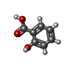  Mass: 138.121 Da / Num. of mol.: 2 / Source method: obtained synthetically / Formula: C7H6O3 Mass: 138.121 Da / Num. of mol.: 2 / Source method: obtained synthetically / Formula: C7H6O3#5: Water | ChemComp-HOH / |  Mass: 18.015 Da / Num. of mol.: 300 / Source method: isolated from a natural source / Formula: H2O Mass: 18.015 Da / Num. of mol.: 300 / Source method: isolated from a natural source / Formula: H2O |
|---|
-Experimental details
-Experiment
| Experiment | Method: X-RAY DIFFRACTION / Number of used crystals: 1 |
|---|
- Sample preparation
| Crystal | Density Matthews: 2.37 Å3/Da / Density % sol: 48.21 % |
|---|---|
| Crystal grow | Temperature: 290 K / Method: vapor diffusion, hanging drop / pH: 6.5 Details: 0.2M magnesium acetate tetrahydrate, 0.1M sodium cacodylate, 20% PEG 8000, pH 6.5, VAPOR DIFFUSION, HANGING DROP, temperature 290K |
-Data collection
| Diffraction | Mean temperature: 100 K |
|---|---|
| Diffraction source | Source: ROTATING ANODE / Type: RIGAKU RUH3R / Wavelength: 1.5418 Å |
| Detector | Type: RIGAKU RAXIS IV / Detector: IMAGE PLATE / Date: Apr 21, 2005 / Details: Rigaku Max Flux mirrors |
| Radiation | Monochromator: Rigaku Max Flux mirrors / Protocol: SINGLE WAVELENGTH / Monochromatic (M) / Laue (L): M / Scattering type: x-ray |
| Radiation wavelength | Wavelength: 1.5418 Å / Relative weight: 1 |
| Reflection | Resolution: 2.1→30 Å / Num. all: 52602 / Num. obs: 49446 / % possible obs: 94 % / Observed criterion σ(F): 2.5 / Observed criterion σ(I): 2.5 / Redundancy: 4.8 % / Biso Wilson estimate: 33.2 Å2 / Rmerge(I) obs: 0.049 / Rsym value: 0.049 / Net I/σ(I): 20.7 |
| Reflection shell | Resolution: 2.1→2.15 Å / Redundancy: 4.7 % / Rmerge(I) obs: 0.299 / Num. unique all: 3453 / Rsym value: 0.299 / % possible all: 88.7 |
- Processing
| Software |
| |||||||||||||||||||||||||||||||||||||||||||||||||||||||||||||||||||||||||||||||||||||||||||||||
|---|---|---|---|---|---|---|---|---|---|---|---|---|---|---|---|---|---|---|---|---|---|---|---|---|---|---|---|---|---|---|---|---|---|---|---|---|---|---|---|---|---|---|---|---|---|---|---|---|---|---|---|---|---|---|---|---|---|---|---|---|---|---|---|---|---|---|---|---|---|---|---|---|---|---|---|---|---|---|---|---|---|---|---|---|---|---|---|---|---|---|---|---|---|---|---|---|
| Refinement | Method to determine structure: MOLECULAR REPLACEMENT Starting model: composite search probe from 1QDL, 1I7Q and 1I1Q Resolution: 2.1→27.37 Å / Cor.coef. Fo:Fc: 0.953 / Cor.coef. Fo:Fc free: 0.923 / SU B: 5.006 / SU ML: 0.135 / Cross valid method: THROUGHOUT / σ(F): 0 / σ(I): 0 / ESU R: 0.232 / ESU R Free: 0.2 / Stereochemistry target values: MAXIMUM LIKELIHOOD / Details: HYDROGENS HAVE BEEN ADDED IN THE RIDING POSITIONS
| |||||||||||||||||||||||||||||||||||||||||||||||||||||||||||||||||||||||||||||||||||||||||||||||
| Solvent computation | Ion probe radii: 0.8 Å / Shrinkage radii: 0.8 Å / VDW probe radii: 1.2 Å / Solvent model: MASK | |||||||||||||||||||||||||||||||||||||||||||||||||||||||||||||||||||||||||||||||||||||||||||||||
| Displacement parameters | Biso mean: 33.63 Å2
| |||||||||||||||||||||||||||||||||||||||||||||||||||||||||||||||||||||||||||||||||||||||||||||||
| Refinement step | Cycle: LAST / Resolution: 2.1→27.37 Å
| |||||||||||||||||||||||||||||||||||||||||||||||||||||||||||||||||||||||||||||||||||||||||||||||
| Refine LS restraints |
| |||||||||||||||||||||||||||||||||||||||||||||||||||||||||||||||||||||||||||||||||||||||||||||||
| LS refinement shell | Resolution: 2.1→2.15 Å / Total num. of bins used: 20
|
