HETEROGEN REGARDING THE LIGAND A37, THE USER SHOULD REFER TO COMPOUND NO. 1 IN THE MAIN REFERENCE ...HETEROGEN REGARDING THE LIGAND A37, THE USER SHOULD REFER TO COMPOUND NO. 1 IN THE MAIN REFERENCE FOR THIS ENTRY, VON GELDERN ET AL. (FIG. 1).
Mass: 377.630 Da / Num. of mol.: 4 / Source method: obtained synthetically / Formula: C13H7Cl3N2O3S
-
Experimental details
-
Experiment
Experiment
Method: X-RAY DIFFRACTION / Number of used crystals: 1
-
Sample preparation
Crystal
Density Matthews: 2.66 Å3/Da / Density % sol: 53.72 %
Crystal grow
Method: vapor diffusion, under oil / pH: 6.5 Details: 0.2 M magnesium acetate, 0.1M sodium cacodylate, 20% PEG8K in the presence of ZMP, pH 6.5, vapor diffusion, under oil
Resolution: 2.95→3.04 Å / Redundancy: 2.4 % / Rmerge(I) obs: 0.304 / Mean I/σ(I) obs: 2.9 / Num. unique all: 2467 / Rsym value: 0.238 / % possible all: 65.7
-
Processing
Software
Name
Version
Classification
CNX
2002
refinement
MAR345
datacollection
HKL-2000
datascaling
CNX
phasing
Refinement
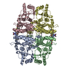
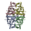
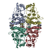
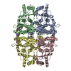
Method to determine structure: MOLECULAR REPLACEMENT / Resolution: 2.95→19.89 Å / Rfactor Rfree error: 0.005 / Data cutoff high absF: 410349.17 / Data cutoff low absF: 0 / Isotropic thermal model: GROUP / Cross valid method: THROUGHOUT / σ(F): 2 / Stereochemistry target values: Engh & Huber Details: Entries 2fhy, 2fie and 2fix characterize the binding mode of benzoxazoles as allosteric inhibitors of human liver FBPase as described in the associated references. These three entries focus ...Details: Entries 2fhy, 2fie and 2fix characterize the binding mode of benzoxazoles as allosteric inhibitors of human liver FBPase as described in the associated references. These three entries focus on the binding mode and interactions in the proximity of the ligands. The medium to low resolution of the crystallographic data (3.3, 2.95 and 2.8 A) and the limited quality and extent of the available data, especially for entry 2fix, should be considered when trying to extract accurate interatomic distances between the ligands and the protein, at certain regions of the protein exposed to solvent.
In the structure databanks used in Yorodumi, some data are registered as the other names, "COVID-19 virus" and "2019-nCoV". Here are the details of the virus and the list of structure data.
Jan 31, 2019. EMDB accession codes are about to change! (news from PDBe EMDB page)
EMDB accession codes are about to change! (news from PDBe EMDB page)
The allocation of 4 digits for EMDB accession codes will soon come to an end. Whilst these codes will remain in use, new EMDB accession codes will include an additional digit and will expand incrementally as the available range of codes is exhausted. The current 4-digit format prefixed with “EMD-” (i.e. EMD-XXXX) will advance to a 5-digit format (i.e. EMD-XXXXX), and so on. It is currently estimated that the 4-digit codes will be depleted around Spring 2019, at which point the 5-digit format will come into force.
The EM Navigator/Yorodumi systems omit the EMD- prefix.
Related info.:Q: What is EMD? / ID/Accession-code notation in Yorodumi/EM Navigator
Yorodumi is a browser for structure data from EMDB, PDB, SASBDB, etc.
This page is also the successor to EM Navigator detail page, and also detail information page/front-end page for Omokage search.
The word "yorodu" (or yorozu) is an old Japanese word meaning "ten thousand". "mi" (miru) is to see.
Related info.:EMDB / PDB / SASBDB / Comparison of 3 databanks / Yorodumi Search / Aug 31, 2016. New EM Navigator & Yorodumi / Yorodumi Papers / Jmol/JSmol / Function and homology information / Changes in new EM Navigator and Yorodumi
 Movie
Movie Controller

 Yorodumi
 Open data
 Basic information
 Components
 Keywords
 Function and homology information
 Homo sapiens (human)
 X-RAY DIFFRACTION /
  Authors
 Citation
 Structure visualization
 Downloads & links
 Other downloads


 PDBj
PDBj
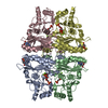
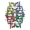
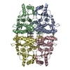
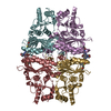
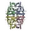
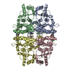
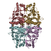
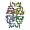
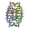
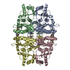
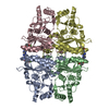
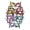
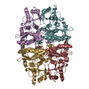
 Assembly
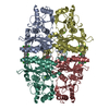


 Mass: 24.305 Da / Num. of mol.: 4 / Source method: obtained synthetically / Formula: Mg
 Mass: 377.630 Da / Num. of mol.: 4 / Source method: obtained synthetically / Formula: C13H7Cl3N2O3S
 Sample preparation
 / Beamline: 17-ID
 Processing