 Movie
Movie Controller

[English] 日本語
 Yorodumi
- PDB-2fix: Structure of human liver FBPase complexed with potent benzoxazole... -
+ Open data

- Basic information
| Entry | Database: PDB / ID: 2fix | ||||||
|---|---|---|---|---|---|---|---|
| Title | Structure of human liver FBPase complexed with potent benzoxazole allosteric inhibitiors | ||||||
 Components Components | Fructose-1,6-bisphosphatase 1 | ||||||
 Keywords Keywords | HYDROLASE / allosteric inhibitors human liver FBPase / benzoxazole / intersubunit allosteric inhibitors of human liver FBPase | ||||||
| Function / homology |  Function and homology information Function and homology informationcellular response to raffinose / cellular hypotonic salinity response / fructose-bisphosphatase / fructose 1,6-bisphosphate 1-phosphatase activity / cellular response to phorbol 13-acetate 12-myristate / fructose 1,6-bisphosphate metabolic process / cellular response to magnesium ion / negative regulation of Ras protein signal transduction / fructose 6-phosphate metabolic process / fructose metabolic process ...cellular response to raffinose / cellular hypotonic salinity response / fructose-bisphosphatase / fructose 1,6-bisphosphate 1-phosphatase activity / cellular response to phorbol 13-acetate 12-myristate / fructose 1,6-bisphosphate metabolic process / cellular response to magnesium ion / negative regulation of Ras protein signal transduction / fructose 6-phosphate metabolic process / fructose metabolic process / monosaccharide binding / Gluconeogenesis / negative regulation of glycolytic process / regulation of gluconeogenesis / cellular hyperosmotic salinity response / AMP binding / cellular response to cAMP / gluconeogenesis / response to nutrient levels / negative regulation of cell growth / cellular response to xenobiotic stimulus / cellular response to insulin stimulus / RNA polymerase II-specific DNA-binding transcription factor binding / negative regulation of transcription by RNA polymerase II / extracellular exosome / metal ion binding / identical protein binding / nucleus / cytoplasm / cytosol Similarity search - Function | ||||||
| Biological species |  Homo sapiens (human) Homo sapiens (human) | ||||||
| Method |  X-RAY DIFFRACTION / SYNCHROTRON / MOLECULAR REPLACEMENT / Resolution: 3.5 Å X-RAY DIFFRACTION / SYNCHROTRON / MOLECULAR REPLACEMENT / Resolution: 3.5 Å | ||||||
 Authors Authors | Abad-Zapatero, C. | ||||||
 Citation Citation | Journal: Bioorg.Med.Chem.Lett. / Year: 2006 Title: Benzoxazole benzenesulfonamides as allosteric inhibitors of fructose-1,6-bisphosphatase. Authors: Lai, C. / Gum, R.J. / Daly, M. / Fry, E.H. / Hutchins, C. / Abad-Zapatero, C. / von Geldern, T.W. #1: Journal: Bioorg.Med.Chem.Lett. / Year: 2006Title: Benzoxazole benzenesulfonamides are novel allosteric inhibitors of fructose-1,6-bisphosphatase with a distinct binding mode. Authors: von Geldern, T.W. / Lai, C. / Gum, R.J. / Daly, M. / Sun, C. / Fry, E.H. / Abad-Zapatero, C. | ||||||
| History |
| ||||||
| Remark 600 | HETEROGEN REGARDING THE LIGAND 870, THE USER SHOULD REFER TO COMPOUND NO. 53 IN THE MAIN REFERENCE ...HETEROGEN REGARDING THE LIGAND 870, THE USER SHOULD REFER TO COMPOUND NO. 53 IN THE MAIN REFERENCE FOR THIS ENTRY, TABLE 3. |
- Structure visualization
| Structure viewer | Molecule: MolmilJmol/JSmol |
|---|
- Downloads & links
-Download
| PDBx/mmCIF format | 2fix.cif.gz | 222.2 KB | Display | PDBx/mmCIF format |
|---|---|---|---|---|
| PDB format | pdb2fix.ent.gz | 172.1 KB | Display | PDB format |
| PDBx/mmJSON format | 2fix.json.gz | Tree view | PDBx/mmJSON format | |
| Others |  Other downloads Other downloads |
-Validation report
| Arichive directory | https://data.pdbj.org/pub/pdb/validation_reports/fi/2fixftp://data.pdbj.org/pub/pdb/validation_reports/fi/2fix | HTTPS FTP |
|---|
-Related structure data
| Related structure data | 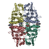 2fieSC S: Starting model for refinement C: citing same article ( |
|---|---|
| Similar structure data |


-Links
 PDBj
PDBj
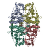
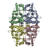
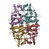
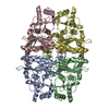
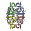
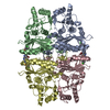
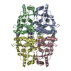
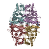
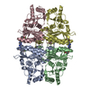
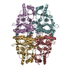
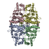
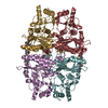
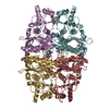
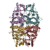
- Assembly
| Deposited unit | 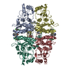
| ||||||||
|---|---|---|---|---|---|---|---|---|---|
| 1 |
| ||||||||
| Unit cell |
| ||||||||
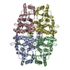
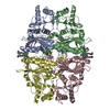
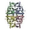
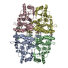
| Details | The biological active unit is the tetramer. Crystal contains four chains (A,D,H,L) and four molecules of the inhibitors (compound here labeled 870 in the asymmetric unit. The biological unit is the same of the other two related entries: 2fhy, 2fie |
-Components
| #1: Protein | Mass: 36859.418 Da / Num. of mol.: 4 Source method: isolated from a genetically manipulated source Source: (gene. exp.) Homo sapiens (human) / Organ: liver / Plasmid: 6His-Xpress-huFBP1-pET100-D-TOPO / Production host:  References: GenBank: 15277851, UniProt: P09467*PLUS, fructose-bisphosphatase #2: Chemical | ChemComp-870 /   Mass: 464.322 Da / Num. of mol.: 4 / Source method: obtained synthetically / Formula: C20H15Cl2N3O4S Mass: 464.322 Da / Num. of mol.: 4 / Source method: obtained synthetically / Formula: C20H15Cl2N3O4S |
|---|
-Experimental details
-Experiment
| Experiment | Method: X-RAY DIFFRACTION / Number of used crystals: 1 |
|---|
- Sample preparation
| Crystal | Density Matthews: 3.05 Å3/Da / Density % sol: 59.71 % |
|---|---|
| Crystal grow | Temperature: 290 K / Method: vapor diffusion, under oil / pH: 6.5 Details: Crystals grown under oil (3:1 parafin:silicon oil). 2 uL+2uL drops. Well solution: 14-18% PEG1000, 0.1 M Tris pH 8.0 Cryo: quick immersion in: 16% PEG1000, 20% PEG400,0.1 M Tris pH 8.0, pH 6. ...Details: Crystals grown under oil (3:1 parafin:silicon oil). 2 uL+2uL drops. Well solution: 14-18% PEG1000, 0.1 M Tris pH 8.0 Cryo: quick immersion in: 16% PEG1000, 20% PEG400,0.1 M Tris pH 8.0, pH 6.5, vapor diffusion, under oil, temperature 290K |
-Data collection
| Diffraction | Mean temperature: 100 K |
|---|---|
| Diffraction source | Source: SYNCHROTRON / Site: APS  / Beamline: 17-BM / Wavelength: 1 Å / Beamline: 17-BM / Wavelength: 1 Å |
| Detector | Type: MARRESEARCH / Detector: CCD / Date: Aug 24, 2004 |
| Radiation | Protocol: SINGLE WAVELENGTH / Monochromatic (M) / Laue (L): M / Scattering type: x-ray |
| Radiation wavelength | Wavelength: 1 Å / Relative weight: 1 |
| Reflection | Resolution: 3.3→20 Å / Num. all: 27761 / Num. obs: 23224 / % possible obs: 88.1 % / Observed criterion σ(F): 1 / Observed criterion σ(I): 1 / Redundancy: 3.5 % / Biso Wilson estimate: 19.5 Å2 / Rmerge(I) obs: 0.245 / Rsym value: 0.245 / Net I/σ(I): 3.5 |
| Reflection shell | Resolution: 3.3→3.42 Å / Redundancy: 2.2 % / Rmerge(I) obs: 0.88 / Mean I/σ(I) obs: 0.69 / Num. unique all: 1651 / Rsym value: 0.88 / % possible all: 60.7 |
- Processing
| Software |
| ||||||||||||||||||||||||||||||||||||||||||||||||||||||||||||
|---|---|---|---|---|---|---|---|---|---|---|---|---|---|---|---|---|---|---|---|---|---|---|---|---|---|---|---|---|---|---|---|---|---|---|---|---|---|---|---|---|---|---|---|---|---|---|---|---|---|---|---|---|---|---|---|---|---|---|---|---|---|
| Refinement | Method to determine structure: MOLECULAR REPLACEMENT Starting model: entry 2fie Resolution: 3.5→19.88 Å / Rfactor Rfree error: 0.009 / Data cutoff high absF: 73686.99 / Data cutoff low absF: 0 / Isotropic thermal model: GROUP / Cross valid method: THROUGHOUT / σ(F): 2 Details: Entries 2fhy, 2fie and 2fix characterize the binding mode of benzoxazoles as allosteric inhibitors of human liver FBPase as described in the associated references. These three entries focus ...Details: Entries 2fhy, 2fie and 2fix characterize the binding mode of benzoxazoles as allosteric inhibitors of human liver FBPase as described in the associated references. These three entries focus on the binding mode and interactions in the proximity of the ligands. The medium to low resolution of the crystallographic data (3.3, 2.95 and 2.8 A) and the limited quality and extent of the available data, especially for entry 2fix, should be considered when trying to extract accurate interatomic distances between the ligands and the protein, at certain regions of the protein exposed to solvent.
| ||||||||||||||||||||||||||||||||||||||||||||||||||||||||||||
| Solvent computation | Solvent model: FLAT MODEL / Bsol: 10 Å2 / ksol: 0.171068 e/Å3 | ||||||||||||||||||||||||||||||||||||||||||||||||||||||||||||
| Displacement parameters | Biso mean: 17.9 Å2
| ||||||||||||||||||||||||||||||||||||||||||||||||||||||||||||
| Refine analyze |
| ||||||||||||||||||||||||||||||||||||||||||||||||||||||||||||
| Refinement step | Cycle: LAST / Resolution: 3.5→19.88 Å
| ||||||||||||||||||||||||||||||||||||||||||||||||||||||||||||
| Refine LS restraints |
| ||||||||||||||||||||||||||||||||||||||||||||||||||||||||||||
| LS refinement shell | Resolution: 3.5→3.66 Å / Rfactor Rfree error: 0.037 / Total num. of bins used: 8
| ||||||||||||||||||||||||||||||||||||||||||||||||||||||||||||
| Xplor file |
|
