 Movie
Movie Controller

[English] 日本語
 Yorodumi
- PDB-2fd5: The crystal structure of a transcriptional regulator from Pseudom... -
+ Open data

- Basic information
| Entry | Database: PDB / ID: 2fd5 | ||||||
|---|---|---|---|---|---|---|---|
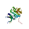
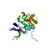
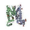
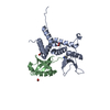
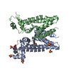
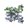
| Title | The crystal structure of a transcriptional regulator from Pseudomonas aeruginosa PAO1 | ||||||
 Components Components | transcriptional regulator | ||||||
 Keywords Keywords | TRANSCRIPTION REGULATOR / Transcriptional regulator / DNA-binding protein / Structural Genomics / PSI / Protein Structure Initiative / Midwest Center for Structural Genomics / MCSG | ||||||
| Function / homology |  Function and homology information Function and homology information | ||||||
| Biological species |   Pseudomonas aeruginosa (bacteria) Pseudomonas aeruginosa (bacteria) | ||||||
| Method |  X-RAY DIFFRACTION / SYNCHROTRON / SAD / Resolution: 1.7 Å X-RAY DIFFRACTION / SYNCHROTRON / SAD / Resolution: 1.7 Å | ||||||
 Authors Authors | Zhang, R. / Skarina, T. / Onopriyenko, O. / Savchenko, A. / Edwards, A. / Joachimiak, A. / Midwest Center for Structural Genomics (MCSG) | ||||||
 Citation Citation | Journal: To be Published Title: The crystal structure of a transcriptional regulator from Pseudomonas aeruginosa PAO1 Authors: Zhang, R. / Skarina, T. / Onopriyenko, O. / Savchenko, A. / Edwards, A. / Joachimiak, A. | ||||||
| History |
|
- Structure visualization
| Structure viewer | Molecule: MolmilJmol/JSmol |
|---|
- Downloads & links
-Download
| PDBx/mmCIF format | 2fd5.cif.gz | 49.7 KB | Display | PDBx/mmCIF format |
|---|---|---|---|---|
| PDB format | pdb2fd5.ent.gz | 35.9 KB | Display | PDB format |
| PDBx/mmJSON format | 2fd5.json.gz | Tree view | PDBx/mmJSON format | |
| Others |  Other downloads Other downloads |
-Validation report
| Arichive directory | https://data.pdbj.org/pub/pdb/validation_reports/fd/2fd5ftp://data.pdbj.org/pub/pdb/validation_reports/fd/2fd5 | HTTPS FTP |
|---|
-Related structure data
| Similar structure data | |
|---|---|
| Other databases |


-Links
 PDBj
PDBj- Assembly
| Deposited unit | 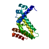
| |||||||||||||||||||||||||||
|---|---|---|---|---|---|---|---|---|---|---|---|---|---|---|---|---|---|---|---|---|---|---|---|---|---|---|---|---|
| 1 | 
| |||||||||||||||||||||||||||
| Unit cell |
| |||||||||||||||||||||||||||
| Components on special symmetry positions |
| |||||||||||||||||||||||||||
| Details | This protein existed as dimer. The second part of the biological assembly is generated by the two fold axis: x-y, -y, -z, plus the translation: 1 2 0 |
-Components
| #1: Protein | Mass: 19200.100 Da / Num. of mol.: 1 Source method: isolated from a genetically manipulated source Source: (gene. exp.) Pseudomonas aeruginosa (bacteria) / Strain: PAO1 / Gene: GI:15598329 / Plasmid: pET15b / Species (production host): Escherichia coli / Production host: |
|---|---|
| #2: Water | ChemComp-HOH /  Mass: 18.015 Da / Num. of mol.: 195 / Source method: isolated from a natural source / Formula: H2O Mass: 18.015 Da / Num. of mol.: 195 / Source method: isolated from a natural source / Formula: H2O |
-Experimental details
-Experiment
| Experiment | Method: X-RAY DIFFRACTION / Number of used crystals: 1 |
|---|
- Sample preparation
| Crystal | Density Matthews: 2.83 Å3/Da / Density % sol: 56.54 % |
|---|---|
| Crystal grow | Temperature: 298 K / Method: vapor diffusion, hanging drop / pH: 5.3 Details: PEG2KMME 30%, NaCitr. 0.1M, NaAcetate 0.2M, Tacsimate 1%, pH 5.3, VAPOR DIFFUSION, HANGING DROP, temperature 298K |
-Data collection
| Diffraction | Mean temperature: 100 K |
|---|---|
| Diffraction source | Source: SYNCHROTRON / Site: APS  / Beamline: 19-ID / Wavelength: 0.9798 Å / Beamline: 19-ID / Wavelength: 0.9798 Å |
| Detector | Type: ADSC QUANTUM 315 / Detector: CCD / Date: Nov 27, 2005 / Details: mirrors |
| Radiation | Monochromator: Si 111 channel / Protocol: SINGLE WAVELENGTH / Monochromatic (M) / Laue (L): M / Scattering type: x-ray |
| Radiation wavelength | Wavelength: 0.9798 Å / Relative weight: 1 |
| Reflection | Resolution: 1.7→61.9 Å / Num. all: 24053 / Num. obs: 23774 / % possible obs: 98.84 % / Observed criterion σ(F): 2 / Observed criterion σ(I): 2 / Redundancy: 12.4 % / Biso Wilson estimate: 22.3 Å2 / Rmerge(I) obs: 0.096 / Net I/σ(I): 27.1 |
| Reflection shell | Resolution: 1.7→1.76 Å / Redundancy: 4.2 % / Rmerge(I) obs: 0.556 / Mean I/σ(I) obs: 1.58 / Num. unique all: 4627 / % possible all: 82.4 |
- Processing
| Software |
| ||||||||||||||||||||||||||||||||||||||||||||||||||||||||||||||||||||||||||||||||||||||||||
|---|---|---|---|---|---|---|---|---|---|---|---|---|---|---|---|---|---|---|---|---|---|---|---|---|---|---|---|---|---|---|---|---|---|---|---|---|---|---|---|---|---|---|---|---|---|---|---|---|---|---|---|---|---|---|---|---|---|---|---|---|---|---|---|---|---|---|---|---|---|---|---|---|---|---|---|---|---|---|---|---|---|---|---|---|---|---|---|---|---|---|---|
| Refinement | Method to determine structure: SAD / Resolution: 1.7→61.9 Å / Cor.coef. Fo:Fc: 0.945 / Cor.coef. Fo:Fc free: 0.929 / SU B: 2.051 / SU ML: 0.069 / Cross valid method: THROUGHOUT / σ(F): 0 / ESU R: 0.108 / ESU R Free: 0.106 Stereochemistry target values: MAXIMUM LIKELIHOOD WITH PHASES Details: HYDROGENS HAVE BEEN ADDED IN THE RIDING POSITIONS
| ||||||||||||||||||||||||||||||||||||||||||||||||||||||||||||||||||||||||||||||||||||||||||
| Solvent computation | Ion probe radii: 0.8 Å / Shrinkage radii: 0.8 Å / VDW probe radii: 1.2 Å / Solvent model: MASK | ||||||||||||||||||||||||||||||||||||||||||||||||||||||||||||||||||||||||||||||||||||||||||
| Displacement parameters | Biso mean: 22.219 Å2
| ||||||||||||||||||||||||||||||||||||||||||||||||||||||||||||||||||||||||||||||||||||||||||
| Refine analyze |
| ||||||||||||||||||||||||||||||||||||||||||||||||||||||||||||||||||||||||||||||||||||||||||
| Refinement step | Cycle: LAST / Resolution: 1.7→61.9 Å
| ||||||||||||||||||||||||||||||||||||||||||||||||||||||||||||||||||||||||||||||||||||||||||
| Refine LS restraints |
| ||||||||||||||||||||||||||||||||||||||||||||||||||||||||||||||||||||||||||||||||||||||||||
| LS refinement shell | Resolution: 1.699→1.743 Å / Total num. of bins used: 20
|
