 Movie
Movie Controller

[English] 日本語
 Yorodumi
- PDB-2f8v: Structure of full length telethonin in complex with the N-terminu... -
+ Open data

- Basic information
| Entry | Database: PDB / ID: 2f8v | ||||||
|---|---|---|---|---|---|---|---|
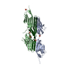
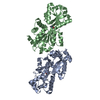
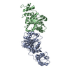
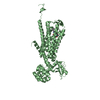
| Title | Structure of full length telethonin in complex with the N-terminus of titin | ||||||
 Components Components |
| ||||||
 Keywords Keywords | CONTRACTILE PROTEIN/CONTRACTILE PROTEIN / Sarcomere / Titin / Z1Z2 / Telethonin / CONTRACTILE PROTEIN-CONTRACTILE PROTEIN COMPLEX | ||||||
| Function / homology |  Function and homology information Function and homology informationFATZ binding / titin Z domain binding / otic vesicle formation / BMP binding / sarcomerogenesis / titin-telethonin complex / structural molecule activity conferring elasticity / skeletal muscle myosin thick filament assembly / telethonin binding / detection of muscle stretch ...FATZ binding / titin Z domain binding / otic vesicle formation / BMP binding / sarcomerogenesis / titin-telethonin complex / structural molecule activity conferring elasticity / skeletal muscle myosin thick filament assembly / telethonin binding / detection of muscle stretch / detection of mechanical stimulus / muscle alpha-actinin binding / : / cardiac myofibril assembly / adult heart development / cardiac muscle hypertrophy / mitotic chromosome condensation / cardiac muscle tissue morphogenesis / Striated Muscle Contraction / cardiac muscle hypertrophy in response to stress / muscle filament sliding / protein kinase regulator activity / actinin binding / M band / I band / cardiac muscle cell development / structural constituent of muscle / sarcomere organization / striated muscle thin filament / skeletal muscle thin filament assembly / skeletal muscle contraction / somitogenesis / striated muscle contraction / cardiac muscle contraction / titin binding / response to muscle stretch / muscle contraction / condensed nuclear chromosome / positive regulation of protein secretion / response to calcium ion / Z disc / actin filament binding / Platelet degranulation / protein-containing complex assembly / protease binding / protein tyrosine kinase activity / protein-macromolecule adaptor activity / molecular adaptor activity / transmembrane transporter binding / calmodulin binding / non-specific serine/threonine protein kinase / protein kinase activity / protein serine kinase activity / protein serine/threonine kinase activity / calcium ion binding / positive regulation of gene expression / protein kinase binding / enzyme binding / protein homodimerization activity / extracellular exosome / extracellular region / ATP binding / identical protein binding / cytosol Similarity search - Function | ||||||
| Biological species |  Homo sapiens (human) Homo sapiens (human) | ||||||
| Method |  X-RAY DIFFRACTION / SYNCHROTRON / MOLECULAR REPLACEMENT / Resolution: 2.75 Å X-RAY DIFFRACTION / SYNCHROTRON / MOLECULAR REPLACEMENT / Resolution: 2.75 Å | ||||||
 Authors Authors | Pinotsis, N. / Petoukhov, M. / Lange, S. / Svergun, D. / Zou, P. / Gautel, M. / Wilmanns, M. | ||||||
 Citation Citation | Journal: J.Struct.Biol. / Year: 2006 Title: Evidence for a dimeric assembly of two titin/telethonin complexes induced by the telethonin C-terminus. Authors: Pinotsis, N. / Petoukhov, M. / Lange, S. / Svergun, D. / Zou, P. / Gautel, M. / Wilmanns, M. | ||||||
| History |
|
- Structure visualization
| Structure viewer | Molecule: MolmilJmol/JSmol |
|---|
- Downloads & links
-Download
| PDBx/mmCIF format | 2f8v.cif.gz | 193.2 KB | Display | PDBx/mmCIF format |
|---|---|---|---|---|
| PDB format | pdb2f8v.ent.gz | 153.6 KB | Display | PDB format |
| PDBx/mmJSON format | 2f8v.json.gz | Tree view | PDBx/mmJSON format | |
| Others |  Other downloads Other downloads |
-Validation report
| Summary document | 2f8v_validation.pdf.gz | 493.7 KB | Display | wwPDB validaton report |
|---|---|---|---|---|
| Full document | 2f8v_full_validation.pdf.gz | 535.9 KB | Display | |
| Data in XML | 2f8v_validation.xml.gz | 38.4 KB | Display | |
| Data in CIF | 2f8v_validation.cif.gz | 52.2 KB | Display | |
| Arichive directory | https://data.pdbj.org/pub/pdb/validation_reports/f8/2f8vftp://data.pdbj.org/pub/pdb/validation_reports/f8/2f8v | HTTPS FTP |
-Related structure data
| Related structure data |  1ya5S S: Starting model for refinement |
|---|---|
| Similar structure data |
-Links
 PDBj
PDBj


- Assembly
| Deposited unit | 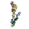
| |||||||||||||||||||||||||||||||||||||||||||||||||||||||||||||||||||||||||||||||||||||||||||||||||||||||||||||||||||||||||||||||||||||||||||||||||||||||||||||||||||||||||||||||||||||||||||||||||||||
|---|---|---|---|---|---|---|---|---|---|---|---|---|---|---|---|---|---|---|---|---|---|---|---|---|---|---|---|---|---|---|---|---|---|---|---|---|---|---|---|---|---|---|---|---|---|---|---|---|---|---|---|---|---|---|---|---|---|---|---|---|---|---|---|---|---|---|---|---|---|---|---|---|---|---|---|---|---|---|---|---|---|---|---|---|---|---|---|---|---|---|---|---|---|---|---|---|---|---|---|---|---|---|---|---|---|---|---|---|---|---|---|---|---|---|---|---|---|---|---|---|---|---|---|---|---|---|---|---|---|---|---|---|---|---|---|---|---|---|---|---|---|---|---|---|---|---|---|---|---|---|---|---|---|---|---|---|---|---|---|---|---|---|---|---|---|---|---|---|---|---|---|---|---|---|---|---|---|---|---|---|---|---|---|---|---|---|---|---|---|---|---|---|---|---|---|---|---|---|
| 1 | 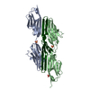
| |||||||||||||||||||||||||||||||||||||||||||||||||||||||||||||||||||||||||||||||||||||||||||||||||||||||||||||||||||||||||||||||||||||||||||||||||||||||||||||||||||||||||||||||||||||||||||||||||||||
| 2 | 
| |||||||||||||||||||||||||||||||||||||||||||||||||||||||||||||||||||||||||||||||||||||||||||||||||||||||||||||||||||||||||||||||||||||||||||||||||||||||||||||||||||||||||||||||||||||||||||||||||||||
| Unit cell |
| |||||||||||||||||||||||||||||||||||||||||||||||||||||||||||||||||||||||||||||||||||||||||||||||||||||||||||||||||||||||||||||||||||||||||||||||||||||||||||||||||||||||||||||||||||||||||||||||||||||
| Noncrystallographic symmetry (NCS) | NCS domain:
NCS domain segments: Refine code: 5
|