[English] 日本語
Yorodumi
- PDB-2f4m: The Mouse PNGase-HR23 Complex Reveals a Complete Remodulation of ... -

+
Open data


ID or keywords:

Loading...

-
Basic information

Entry
Database: PDB / ID: 2f4m
TitleThe Mouse PNGase-HR23 Complex Reveals a Complete Remodulation of the Protein-Protein Interface Compared to its Yeast Orthologs
Components
  • UV excision repair protein RAD23 homolog B
  • peptide N-glycanase
KeywordsHYDROLASE / Glycoproteins / Ubiquitin-dependent protein degradation / Nucleotide excision repair / Peptide:N-glycanase / Transglutaminase
Function / homology
Function and homology information


peptide-N4-(N-acetyl-beta-glucosaminyl)asparagine amidase / peptide-N4-(N-acetyl-beta-glucosaminyl)asparagine amidase activity / DNA Damage Recognition in GG-NER / XPC complex / Josephin domain DUBs / N-glycan trimming in the ER and Calnexin/Calreticulin cycle / Formation of Incision Complex in GG-NER / glycoprotein catabolic process / DNA damage sensor activity / regulation of proteasomal ubiquitin-dependent protein catabolic process ...peptide-N4-(N-acetyl-beta-glucosaminyl)asparagine amidase / peptide-N4-(N-acetyl-beta-glucosaminyl)asparagine amidase activity / DNA Damage Recognition in GG-NER / XPC complex / Josephin domain DUBs / N-glycan trimming in the ER and Calnexin/Calreticulin cycle / Formation of Incision Complex in GG-NER / glycoprotein catabolic process / DNA damage sensor activity / regulation of proteasomal ubiquitin-dependent protein catabolic process / cellular response to interleukin-7 / proteasome binding / protein quality control for misfolded or incompletely synthesized proteins / polyubiquitin modification-dependent protein binding / embryonic organ development / proteasome complex / ubiquitin binding / nucleotide-excision repair / spermatogenesis / proteasome-mediated ubiquitin-dependent protein catabolic process / collagen-containing extracellular matrix / RNA polymerase II-specific DNA-binding transcription factor binding / damaged DNA binding / transcription cis-regulatory region binding / RNA polymerase II cis-regulatory region sequence-specific DNA binding / DNA damage response / nucleoplasm / nucleus / metal ion binding / cytoplasm / cytosol
Similarity search - Function
Peptide N glycanase, PAW domain / PAW domain superfamily / PNGase C-terminal domain, mannose-binding module PAW / PAW domain profile. / domain present in PNGases and other hypothetical proteins / XPC-binding domain / C8orf32 fold - #30 / C8orf32 fold / RAD23A/RAD23B, UBA1 domain / UV excision repair protein Rad23 ...Peptide N glycanase, PAW domain / PAW domain superfamily / PNGase C-terminal domain, mannose-binding module PAW / PAW domain profile. / domain present in PNGases and other hypothetical proteins / XPC-binding domain / C8orf32 fold - #30 / C8orf32 fold / RAD23A/RAD23B, UBA1 domain / UV excision repair protein Rad23 / XPC-binding domain / XPC-binding domain superfamily / XPC-binding domain / PUB-like domain superfamily / PUB domain / PUB domain / domain in protein kinases, N-glycanases and other nuclear proteins / Transglutaminase-like superfamily / Transglutaminase/protease-like homologues / Transglutaminase-like / Heat shock chaperonin-binding / Heat shock chaperonin-binding motif. / N-terminal domain of TfIIb - #10 / UBA/TS-N domain / Ubiquitin associated domain / Ubiquitin-associated domain / Ubiquitin-associated domain (UBA) profile. / N-terminal domain of TfIIb / UBA-like superfamily / Single Sheet / Papain-like cysteine peptidase superfamily / Galactose-binding-like domain superfamily / Ubiquitin family / Ubiquitin homologues / Ubiquitin domain profile. / Ubiquitin-like domain / Arc Repressor Mutant, subunit A / Ubiquitin-like domain superfamily / Roll / Orthogonal Bundle / Mainly Beta / Mainly Alpha / Alpha Beta
Similarity search - Domain/homology
UV excision repair protein RAD23 homolog B / Peptide-N(4)-(N-acetyl-beta-glucosaminyl)asparagine amidase
Similarity search - Component
Biological speciesMus musculus (house mouse)
MethodX-RAY DIFFRACTION / MIR / Resolution: 1.85 Å
AuthorsZhao, G. / Zhou, X. / Wang, L. / Kisker, C. / Lennarz, W.J. / Schindelin, H.
CitationJournal: J.Biol.Chem. / Year: 2006
Title: Structure of the mouse peptide N-glycanase-HR23 complex suggests co-evolution of the endoplasmic reticulum-associated degradation and DNA repair pathways.
Authors: Zhao, G. / Zhou, X. / Wang, L. / Li, G. / Kisker, C. / Lennarz, W.J. / Schindelin, H.
History
DepositionNov 23, 2005Deposition site: RCSB / Processing site: RCSB
Revision 1.0Mar 7, 2006Provider: repository / Type: Initial release
Revision 1.1May 1, 2008Group: Version format compliance
Revision 1.2Jul 13, 2011Group: Advisory / Version format compliance

-
Structure visualization

Structure viewerMolecule:
MolmilJmol/JSmol

Downloads & links

-
Assembly

Deposited unit
A: peptide N-glycanase
B: UV excision repair protein RAD23 homolog B
hetero molecules


Theoretical massNumber of molelcules
Total (without water)42,1534
Polymers42,0522
Non-polymers1012
Water4,594255
1


  • Idetical with deposited unit
  • defined by author&software
TypeNameSymmetry operationNumber
identity operation1_555x,y,z1
Buried area1920 Å2
ΔGint-30 kcal/mol
Surface area18340 Å2
MethodPISA
2
A: peptide N-glycanase
B: UV excision repair protein RAD23 homolog B
hetero molecules

A: peptide N-glycanase
B: UV excision repair protein RAD23 homolog B
hetero molecules


Theoretical massNumber of molelcules
Total (without water)84,3068
Polymers84,1044
Non-polymers2024
Water724
TypeNameSymmetry operationNumber
identity operation1_555x,y,z1
crystal symmetry operation2_656-x+1,y,-z+11
Buried area5670 Å2
ΔGint-68 kcal/mol
Surface area34840 Å2
MethodPISA
Unit cell
Length a, b, c (Å)98.225, 52.297, 82.548
Angle α, β, γ (deg.)90.00, 115.33, 90.00
Int Tables number5
Space group name H-MC121

-
Components

#1: Protein peptide N-glycanase


Mass: 34866.730 Da / Num. of mol.: 1 / Fragment: catalytic domain, residues 164-450
Source method: isolated from a genetically manipulated source
Source: (gene. exp.) Mus musculus (house mouse) / Plasmid: pET21b / Production host: Escherichia coli (E. coli) / Strain (production host): BL21-CodonPlus-RIL
References: UniProt: Q9JI78, peptide-N4-(N-acetyl-beta-glucosaminyl)asparagine amidase
#2: Protein UV excision repair protein RAD23 homolog B / mHR23B / XP-C repair complementing complex 58 kDa protein / p58


Mass: 7185.165 Da / Num. of mol.: 1 / Fragment: XPCB domain, residues 273-332
Source method: isolated from a genetically manipulated source
Source: (gene. exp.) Mus musculus (house mouse) / Gene: Rad23b, Mhr23b / Plasmid: pTYB1 / Production host: Escherichia coli (E. coli) / Strain (production host): BL21-CodonPlus-RIL / References: UniProt: P54728
#3: Chemical ChemComp-ZN / ZINC ION


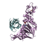
Mass: 65.409 Da / Num. of mol.: 1 / Source method: obtained synthetically / Formula: Zn
#4: Chemical ChemComp-CL / CHLORIDE ION


Mass: 35.453 Da / Num. of mol.: 1 / Source method: obtained synthetically / Formula: Cl
#5: Water ChemComp-HOH / water


Mass: 18.015 Da / Num. of mol.: 255 / Source method: isolated from a natural source / Formula: H2O

-
Experimental details

-
Experiment

ExperimentMethod: X-RAY DIFFRACTION / Number of used crystals: 5

-
Sample preparation

CrystalDensity Matthews: 2.27 Å3/Da / Density % sol: 45.91 %
Crystal growTemperature: 291 K / pH: 8.5
Details: 0.1 M Tris-HCl, pH 8.5, 28-32% PEG4000, 0.2 M sodium acetate, VAPOR DIFFUSION, HANGING DROP, temperature 291K, pH 8.50

-
Data collection

DiffractionMean temperature: 100 K
Diffraction sourceSource: ROTATING ANODE / Type: RIGAKU / Wavelength: 1.5418
DetectorType: RIGAKU RAXIS IV / Detector: IMAGE PLATE / Date: Apr 23, 2005
RadiationMonochromator: GRAPHITE / Protocol: SINGLE WAVELENGTH / Monochromatic (M) / Laue (L): M / Scattering type: x-ray
Radiation wavelengthWavelength: 1.5418 Å / Relative weight: 1
ReflectionResolution: 1.85→50 Å / Num. obs: 31216 / % possible obs: 95.9 % / Observed criterion σ(I): 0 / Redundancy: 4.5 % / Biso Wilson estimate: 42 Å2 / Rsym value: 0.051 / Net I/σ(I): 15.5
Reflection shellResolution: 1.85→1.91 Å / Mean I/σ(I) obs: 2 / Rsym value: 0.4 / % possible all: 20.9

-
Processing

Software
NameVersionClassification
REFMAC5.2.0005refinement
DENZOdata reduction
SCALEPACKdata scaling
SOLVEphasing
RefinementMethod to determine structure: MIR / Resolution: 1.85→20 Å / Cor.coef. Fo:Fc: 0.966 / Cor.coef. Fo:Fc free: 0.94 / SU B: 7.672 / SU ML: 0.122 / TLS residual ADP flag: LIKELY RESIDUAL / Cross valid method: THROUGHOUT / σ(F): 2 / ESU R: 0.155 / ESU R Free: 0.15 / Stereochemistry target values: MAXIMUM LIKELIHOOD / Details: HYDROGENS HAVE BEEN ADDED IN THE RIDING POSITIONS
RfactorNum. reflection% reflectionSelection details
Rfree0.239 1579 5.1 %RANDOM
Rwork0.186 ---
obs0.189 29636 96 %-
all-40360 --
Solvent computationIon probe radii: 0.8 Å / Shrinkage radii: 0.8 Å / VDW probe radii: 1.2 Å / Solvent model: MASK
Displacement parametersBiso mean: 42.62 Å2
Baniso -1Baniso -2Baniso -3
1-1.55 Å20 Å20.23 Å2
2--1.07 Å20 Å2
3----2.43 Å2
Refinement stepCycle: LAST / Resolution: 1.85→20 Å
ProteinNucleic acidLigandSolventTotal
Num. atoms2953 0 2 255 3210
Refine LS restraints
Refine-IDTypeDev idealDev ideal targetNumber
X-RAY DIFFRACTIONr_bond_refined_d0.0160.0213023
X-RAY DIFFRACTIONr_bond_other_d0.0020.022685
X-RAY DIFFRACTIONr_angle_refined_deg1.5471.9454086
X-RAY DIFFRACTIONr_angle_other_deg0.91536274
X-RAY DIFFRACTIONr_dihedral_angle_1_deg6.9095354
X-RAY DIFFRACTIONr_dihedral_angle_2_deg38.56124.146164
X-RAY DIFFRACTIONr_dihedral_angle_3_deg17.3715553
X-RAY DIFFRACTIONr_dihedral_angle_4_deg21.9251525
X-RAY DIFFRACTIONr_chiral_restr0.0930.2432
X-RAY DIFFRACTIONr_gen_planes_refined0.0060.023334
X-RAY DIFFRACTIONr_gen_planes_other0.0010.02614
X-RAY DIFFRACTIONr_nbd_refined0.2140.2642
X-RAY DIFFRACTIONr_nbd_other0.1960.22797
X-RAY DIFFRACTIONr_nbtor_refined0.1730.21377
X-RAY DIFFRACTIONr_nbtor_other0.0840.21684
X-RAY DIFFRACTIONr_xyhbond_nbd_refined0.2590.2178
X-RAY DIFFRACTIONr_xyhbond_nbd_other
X-RAY DIFFRACTIONr_metal_ion_refined
X-RAY DIFFRACTIONr_metal_ion_other
X-RAY DIFFRACTIONr_symmetry_vdw_refined0.1530.222
X-RAY DIFFRACTIONr_symmetry_vdw_other0.1680.257
X-RAY DIFFRACTIONr_symmetry_hbond_refined0.3340.219
X-RAY DIFFRACTIONr_symmetry_hbond_other
X-RAY DIFFRACTIONr_symmetry_metal_ion_refined
X-RAY DIFFRACTIONr_symmetry_metal_ion_other
X-RAY DIFFRACTIONr_mcbond_it1.011.52333
X-RAY DIFFRACTIONr_mcbond_other0.2491.5715
X-RAY DIFFRACTIONr_mcangle_it1.21422881
X-RAY DIFFRACTIONr_scbond_it2.00931443
X-RAY DIFFRACTIONr_scangle_it2.7934.51205
X-RAY DIFFRACTIONr_rigid_bond_restr
X-RAY DIFFRACTIONr_sphericity_free
X-RAY DIFFRACTIONr_sphericity_bonded
LS refinement shellResolution: 1.85→1.9 Å / Total num. of bins used: 20
RfactorNum. reflection% reflection
Rfree0.357 68 -
Rwork0.28 1474 -
obs--65.64 %
Refinement TLS params.

Method: refined / Refine-ID: X-RAY DIFFRACTION

IDL112)L122)L132)L222)L232)L332)S11 (Å °)S12 (Å °)S13 (Å °)S21 (Å °)S22 (Å °)S23 (Å °)S31 (Å °)S32 (Å °)S33 (Å °)T112)T122)T132)T222)T232)T332)Origin x (Å)Origin y (Å)Origin z (Å)
18.69481.0772-4.7865.4009-0.63037.151-0.470.2366-0.7672-0.2132-0.0814-0.44451.06020.21780.55140.01270.01090.1361-0.1252-0.027-0.170543.53342.17597.4902
23.7935-0.7945-1.85053.26830.00785.21960.21540.24740.5588-0.17510.00660.0275-0.6417-0.352-0.222-0.16020.02820.0012-0.2330.0401-0.239527.818519.480619.2027
39.4823-3.5453-8.60584.40193.296721.1726-0.25490.7513-0.582-0.05930.02630.49180.5479-0.72780.2285-0.15320.10960.010.07530.0153-0.01594.034721.268537.64
Refinement TLS group
IDRefine-IDRefine TLS-IDAuth asym-IDLabel asym-IDAuth seq-IDLabel seq-ID
1X-RAY DIFFRACTION1BB273 - 3331 - 61
2X-RAY DIFFRACTION2AA164 - 2411 - 78
3X-RAY DIFFRACTION2AA292 - 458129 - 295
4X-RAY DIFFRACTION3AA242 - 29179 - 128
5X-RAY DIFFRACTION3AC5011

+
About Yorodumi

-
News

-
Feb 9, 2022. New format data for meta-information of EMDB entries

New format data for meta-information of EMDB entries

  • Version 3 of the EMDB header file is now the official format.
  • The previous official version 1.9 will be removed from the archive.

Related info.:EMDB header

External links:wwPDB to switch to version 3 of the EMDB data model

-
Aug 12, 2020. Covid-19 info

Covid-19 info

URL: https://pdbjlvh1.pdbj.org/emnavi/covid19.php

New page: Covid-19 featured information page in EM Navigator.

Related info.:Covid-19 info / Mar 5, 2020. Novel coronavirus structure data

+
Mar 5, 2020. Novel coronavirus structure data

Novel coronavirus structure data

Related info.:Yorodumi Speices / Aug 12, 2020. Covid-19 info

External links:COVID-19 featured content - PDBj / Molecule of the Month (242):Coronavirus Proteases

+
Jan 31, 2019. EMDB accession codes are about to change! (news from PDBe EMDB page)

EMDB accession codes are about to change! (news from PDBe EMDB page)

  • The allocation of 4 digits for EMDB accession codes will soon come to an end. Whilst these codes will remain in use, new EMDB accession codes will include an additional digit and will expand incrementally as the available range of codes is exhausted. The current 4-digit format prefixed with “EMD-” (i.e. EMD-XXXX) will advance to a 5-digit format (i.e. EMD-XXXXX), and so on. It is currently estimated that the 4-digit codes will be depleted around Spring 2019, at which point the 5-digit format will come into force.
  • The EM Navigator/Yorodumi systems omit the EMD- prefix.

Related info.:Q: What is EMD? / ID/Accession-code notation in Yorodumi/EM Navigator

External links:EMDB Accession Codes are Changing Soon! / Contact to PDBj

+
Jul 12, 2017. Major update of PDB

Major update of PDB

  • wwPDB released updated PDB data conforming to the new PDBx/mmCIF dictionary.
  • This is a major update changing the version number from 4 to 5, and with Remediation, in which all the entries are updated.
  • In this update, many items about electron microscopy experimental information are reorganized (e.g. em_software).
  • Now, EM Navigator and Yorodumi are based on the updated data.

External links:wwPDB Remediation / Enriched Model Files Conforming to OneDep Data Standards Now Available in the PDB FTP Archive

-
Yorodumi

Thousand views of thousand structures

  • Yorodumi is a browser for structure data from EMDB, PDB, SASBDB, etc.
  • This page is also the successor to EM Navigator detail page, and also detail information page/front-end page for Omokage search.
  • The word "yorodu" (or yorozu) is an old Japanese word meaning "ten thousand". "mi" (miru) is to see.

Related info.:EMDB / PDB / SASBDB / Comparison of 3 databanks / Yorodumi Search / Aug 31, 2016. New EM Navigator & Yorodumi / Yorodumi Papers / Jmol/JSmol / Function and homology information / Changes in new EM Navigator and Yorodumi

Read more