 Movie
Movie Controller


+ Open data

- Basic information
| Entry | Database: PDB / ID: 3vh4 | ||||||
|---|---|---|---|---|---|---|---|
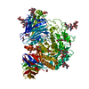
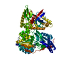
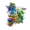
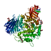
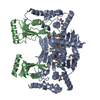
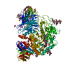
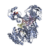
| Title | Crystal structure of Atg7CTD-Atg8-MgATP complex | ||||||
 Components Components |
| ||||||
 Keywords Keywords | METAL BINDING PROTEIN/PROTEIN TRANSPORT / autophagy / E1 / zinc binding / METAL BINDING PROTEIN-PROTEIN TRANSPORT complex | ||||||
| Function / homology |  Function and homology information Function and homology informationCvt vesicle membrane / Atg12 activating enzyme activity / Atg8 activating enzyme activity / TBC/RABGAPs / Receptor Mediated Mitophagy / protein modification by small protein conjugation / extrinsic component of phagophore assembly site membrane / vacuole-isolation membrane contact site / regulation of membrane invagination / protein targeting to vacuole involved in autophagy ...Cvt vesicle membrane / Atg12 activating enzyme activity / Atg8 activating enzyme activity / TBC/RABGAPs / Receptor Mediated Mitophagy / protein modification by small protein conjugation / extrinsic component of phagophore assembly site membrane / vacuole-isolation membrane contact site / regulation of membrane invagination / protein targeting to vacuole involved in autophagy / Macroautophagy / lipid droplet formation / cytoplasm to vacuole targeting by the Cvt pathway / nucleophagy / protein localization to phagophore assembly site / autophagy of mitochondrion / piecemeal microautophagy of the nucleus / phosphatidylethanolamine binding / fungal-type vacuole membrane / phagophore assembly site / reticulophagy / cellular response to nitrogen starvation / protein-containing complex localization / Antigen processing: Ubiquitination & Proteasome degradation / autophagosome membrane / regulation of macroautophagy / autophagosome maturation / endoplasmic reticulum to Golgi vesicle-mediated transport / autophagosome assembly / mitophagy / Neutrophil degranulation / lipid droplet / autophagosome / macroautophagy / autophagy / mitochondrial membrane / protein tag activity / membrane fusion / mitochondrion / membrane / identical protein binding / cytoplasm / cytosol Similarity search - Function | ||||||
| Biological species |  | ||||||
| Method |  X-RAY DIFFRACTION / SYNCHROTRON / MOLECULAR REPLACEMENT / Resolution: 2.65 Å X-RAY DIFFRACTION / SYNCHROTRON / MOLECULAR REPLACEMENT / Resolution: 2.65 Å | ||||||
 Authors Authors | Noda, N.N. / Satoo, K. / Inagaki, F. | ||||||
 Citation Citation | Journal: Mol.Cell / Year: 2011 Title: Structural basis of Atg8 activation by a homodimeric E1, Atg7. Authors: Noda, N.N. / Satoo, K. / Fujioka, Y. / Kumeta, H. / Ogura, K. / Nakatogawa, H. / Ohsumi, Y. / Inagaki, F. | ||||||
| History |
|
- Structure visualization
| Structure viewer | Molecule: MolmilJmol/JSmol |
|---|
- Downloads & links
-Download
| PDBx/mmCIF format | 3vh4.cif.gz | 98.8 KB | Display | PDBx/mmCIF format |
|---|---|---|---|---|
| PDB format | pdb3vh4.ent.gz | 72.6 KB | Display | PDB format |
| PDBx/mmJSON format | 3vh4.json.gz | Tree view | PDBx/mmJSON format | |
| Others |  Other downloads Other downloads |
-Validation report
| Arichive directory | https://data.pdbj.org/pub/pdb/validation_reports/vh/3vh4ftp://data.pdbj.org/pub/pdb/validation_reports/vh/3vh4 | HTTPS FTP |
|---|
-Related structure data
| Related structure data | 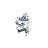 2li5C  3vh1C  3vh2C 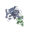 3vh3SC C: citing same article ( S: Starting model for refinement |
|---|---|
| Similar structure data |


-Links
 PDBj
PDBj


- Assembly
| Deposited unit | 
| ||||||||
|---|---|---|---|---|---|---|---|---|---|
| 1 | 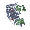
| ||||||||
| Unit cell |
|
-Components
| #1: Protein | Mass: 38150.859 Da / Num. of mol.: 1 / Fragment: C-terminal domain (UNP RESIDUES 295-630) Source method: isolated from a genetically manipulated source Source: (gene. exp.) Strain: S288c / Gene: ATG7, APG7, CVT2, YHR171W / Plasmid: pGEX6P / Production host:  |
|---|---|
| #2: Protein | Mass: 13751.865 Da / Num. of mol.: 1 / Mutation: K26P Source method: isolated from a genetically manipulated source Source: (gene. exp.) Strain: S288c / Gene: ATG8, APG8, AUT7, CVT5, YBL078C, YBL0732 / Plasmid: pGEX6P / Production host: |
| #3: Chemical | ChemComp-ZN /   Mass: 65.409 Da / Num. of mol.: 1 / Source method: obtained synthetically / Formula: Zn Mass: 65.409 Da / Num. of mol.: 1 / Source method: obtained synthetically / Formula: Zn |
| #4: Chemical | ChemComp-ATP /   Mass: 507.181 Da / Num. of mol.: 1 / Source method: obtained synthetically / Formula: C10H16N5O13P3 / Comment: ATP, energy-carrying molecule*YM Mass: 507.181 Da / Num. of mol.: 1 / Source method: obtained synthetically / Formula: C10H16N5O13P3 / Comment: ATP, energy-carrying molecule*YM |
| #5: Chemical | ChemComp-MG /   Mass: 24.305 Da / Num. of mol.: 1 / Source method: obtained synthetically / Formula: Mg Mass: 24.305 Da / Num. of mol.: 1 / Source method: obtained synthetically / Formula: Mg |
-Experimental details
-Experiment
| Experiment | Method: X-RAY DIFFRACTION / Number of used crystals: 1 |
|---|
- Sample preparation
| Crystal | Density Matthews: 2.53 Å3/Da / Density % sol: 51.43 % |
|---|---|
| Crystal grow | Temperature: 293 K / Method: vapor diffusion, sitting drop / pH: 7 Details: 10% PEGMME 5000, 5% tacsimate, 0.1M HEPES, pH 7.0, VAPOR DIFFUSION, SITTING DROP, temperature 293K |
-Data collection
| Diffraction | Mean temperature: 90 K |
|---|---|
| Diffraction source | Source: SYNCHROTRON / Site: Photon Factory  / Beamline: BL-5A / Wavelength: 1 Å / Beamline: BL-5A / Wavelength: 1 Å |
| Detector | Type: ADSC QUANTUM 315 / Detector: CCD / Date: Feb 2, 2011 |
| Radiation | Protocol: SINGLE WAVELENGTH / Monochromatic (M) / Laue (L): M / Scattering type: x-ray |
| Radiation wavelength | Wavelength: 1 Å / Relative weight: 1 |
| Reflection | Resolution: 2.65→50 Å / Num. all: 16447 / Num. obs: 16404 / % possible obs: 99.7 % / Observed criterion σ(F): 0 / Observed criterion σ(I): -3 / Redundancy: 22.7 % / Biso Wilson estimate: 31.4 Å2 / Rmerge(I) obs: 0.07 / Net I/σ(I): 73.1 |
| Reflection shell | Resolution: 2.65→2.7 Å / Redundancy: 14.1 % / Rmerge(I) obs: 0.458 / Mean I/σ(I) obs: 9.2 / % possible all: 100 |
- Processing
| Software |
| ||||||||||||||||||||||||||||||||||||||||||||||||||||||||||||||||||||||||||||||||
|---|---|---|---|---|---|---|---|---|---|---|---|---|---|---|---|---|---|---|---|---|---|---|---|---|---|---|---|---|---|---|---|---|---|---|---|---|---|---|---|---|---|---|---|---|---|---|---|---|---|---|---|---|---|---|---|---|---|---|---|---|---|---|---|---|---|---|---|---|---|---|---|---|---|---|---|---|---|---|---|---|---|
| Refinement | Method to determine structure: MOLECULAR REPLACEMENT Starting model: PDB ENTRY 3VH3 Resolution: 2.65→33.04 Å / Rfactor Rfree error: 0.007 / Data cutoff high absF: 103999 / Data cutoff low absF: 0 / Isotropic thermal model: RESTRAINED / Cross valid method: THROUGHOUT / σ(F): 0 / Stereochemistry target values: Engh & Huber / Details: BULK SOLVENT MODEL USED
| ||||||||||||||||||||||||||||||||||||||||||||||||||||||||||||||||||||||||||||||||
| Solvent computation | Solvent model: FLAT MODEL / Bsol: 41.8378 Å2 / ksol: 0.32 e/Å3 | ||||||||||||||||||||||||||||||||||||||||||||||||||||||||||||||||||||||||||||||||
| Displacement parameters | Biso mean: 82.6 Å2
| ||||||||||||||||||||||||||||||||||||||||||||||||||||||||||||||||||||||||||||||||
| Refine analyze |
| ||||||||||||||||||||||||||||||||||||||||||||||||||||||||||||||||||||||||||||||||
| Refinement step | Cycle: LAST / Resolution: 2.65→33.04 Å
| ||||||||||||||||||||||||||||||||||||||||||||||||||||||||||||||||||||||||||||||||
| Refine LS restraints |
| ||||||||||||||||||||||||||||||||||||||||||||||||||||||||||||||||||||||||||||||||
| LS refinement shell | Resolution: 2.65→2.82 Å / Rfactor Rfree error: 0.027 / Total num. of bins used: 6
| ||||||||||||||||||||||||||||||||||||||||||||||||||||||||||||||||||||||||||||||||
| Xplor file |
|
