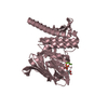
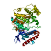
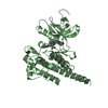
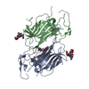
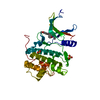
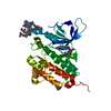
Entry Database : PDB / ID : 2f4jTitle Structure of the Kinase Domain of an Imatinib-Resistant Abl Mutant in Complex with the Aurora Kinase Inhibitor VX-680 Proto-oncogene tyrosine-protein kinase ABL1 Keywords / / / Function / homology Function Domain/homology Component
/ / / / / / / / / / / / / / / / / / / / / / / / / / / / / / / / / / / / / / / / / / / / / / / / / / / / / / / / / / / / / / / / / / / / / / / / / / / / / / / / / / / / / / / / / / / / / / / / / / / / / / / / / / / / / / / / / / / / / / / / / / / / / / / / / / / / / / / / / / / / / / / / / / / / / / Biological species Homo sapiens (human)Method / / / Resolution : 1.91 Å Authors Young, M.A. / Shah, N.P. / Chao, L.H. / Zarrinkar, P. / Sawyers, P. / Kuriyan, J. Journal : Cancer Res. / Year : 2006Title : Structure of the kinase domain of an imatinib-resistant Abl mutant in complex with the Aurora kinase inhibitor VX-680.Authors : Young, M.A. / Shah, N.P. / Chao, L.H. / Seeliger, M. / Milanov, Z.V. / Biggs, W.H. / Treiber, D.K. / Patel, H.K. / Zarrinkar, P.P. / Lockhart, D.J. / Sawyers, C.L. / Kuriyan, J. History Deposition Nov 23, 2005 Deposition site / Processing site Revision 1.0 Jan 24, 2006 Provider / Type Revision 1.1 May 1, 2008 Group Revision 1.2 Jul 13, 2011 Group Revision 1.3 Oct 18, 2017 Group / Category Item _software.classification / _software.contact_author ... _software.classification / _software.contact_author / _software.contact_author_email / _software.date / _software.language / _software.location / _software.name / _software.type / _software.version Revision 1.4 Oct 20, 2021 Group / Derived calculations / Category / struct_ref_seq_dif / struct_siteItem _database_2.pdbx_DOI / _database_2.pdbx_database_accession ... _database_2.pdbx_DOI / _database_2.pdbx_database_accession / _struct_ref_seq_dif.details / _struct_site.pdbx_auth_asym_id / _struct_site.pdbx_auth_comp_id / _struct_site.pdbx_auth_seq_id Revision 1.5 Aug 23, 2023 Group / Refinement descriptionCategory / chem_comp_bond / pdbx_initial_refinement_model
Show all Show less
 Movie
Movie Controller

 Yorodumi
 Open data
 Basic information
 Components
 Keywords
 Function and homology information
 Homo sapiens (human)
 X-RAY DIFFRACTION /
  Authors
 Citation
 Structure visualization
 Downloads & links
 Other downloads


 PDBj
PDBj


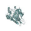
 Assembly


 Mass: 464.586 Da / Num. of mol.: 1 / Source method: obtained synthetically / Formula: C23H28N8OS
 Mass: 18.015 Da / Num. of mol.: 211 / Source method: isolated from a natural source / Formula: H2O
 Sample preparation
 / Beamline: 8.2.1 / Wavelength: 1 Å
 Processing