 Movie
Movie Controller

[English] 日本語
 Yorodumi
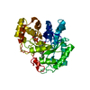
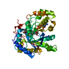
- PDB-2f38: Crystal structure of prostaglandin F synathase containing bimatoprost -
+ Open data

- Basic information
| Entry | Database: PDB / ID: 2f38 | ||||||
|---|---|---|---|---|---|---|---|
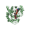
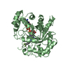
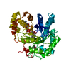
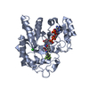
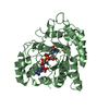
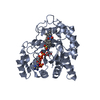
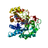
| Title | Crystal structure of prostaglandin F synathase containing bimatoprost | ||||||
 Components Components | Aldo-keto reductase family 1 member C3 | ||||||
 Keywords Keywords | OXIDOREDUCTASE / Prostaglandin F synthase / AKR1C3 / PGF2alpha fromation / PGH2 / bimatoprost / catalytic mechanism | ||||||
| Function / homology |  Function and homology information Function and homology informationprostaglandin-F synthase / testosterone 17beta-dehydrogenase (NADP+) / cellular response to prostaglandin stimulus / cellular response to corticosteroid stimulus / prostaglandin F synthase activity / macromolecule metabolic process / 3beta(or 20alpha)-hydroxysteroid dehydrogenase / prostaglandin D2 11-ketoreductase activity / ketoreductase activity / negative regulation of retinoic acid biosynthetic process ...prostaglandin-F synthase / testosterone 17beta-dehydrogenase (NADP+) / cellular response to prostaglandin stimulus / cellular response to corticosteroid stimulus / prostaglandin F synthase activity / macromolecule metabolic process / 3beta(or 20alpha)-hydroxysteroid dehydrogenase / prostaglandin D2 11-ketoreductase activity / ketoreductase activity / negative regulation of retinoic acid biosynthetic process / 5-alpha-androstane-3-beta,17-beta-diol dehydrogenase (NADP+) activity / 15-hydroxyprostaglandin-D dehydrogenase (NADP+) activity / farnesol catabolic process / 3alpha-hydroxysteroid 3-dehydrogenase / cellular response to jasmonic acid stimulus / Delta4-3-oxosteroid 5beta-reductase activity / 3alpha(17beta)-hydroxysteroid dehydrogenase (NAD+) / : / testosterone dehydrogenase (NADP+) activity / 3-alpha-hydroxysteroid 3-dehydrogenase [NAD(P)+] activity / androsterone dehydrogenase [NAD(P)+] activity / 3alpha(or 20beta)-hydroxysteroid dehydrogenase / RA biosynthesis pathway / regulation of testosterone biosynthetic process / ketosteroid monooxygenase activity / testosterone biosynthetic process / androstan-3-alpha,17-beta-diol dehydrogenase (NAD+) activity / Synthesis of bile acids and bile salts via 24-hydroxycholesterol / testosterone dehydrogenase (NAD+) activity / regulation of retinoic acid receptor signaling pathway / cellular response to prostaglandin D stimulus / prostanoid biosynthetic process / progesterone metabolic process / 17beta-estradiol 17-dehydrogenase / retinal metabolic process / estradiol 17-beta-dehydrogenase [NAD(P)+] activity / Synthesis of Prostaglandins (PG) and Thromboxanes (TX) / all-trans-retinol dehydrogenase (NAD+) activity / alcohol dehydrogenase (NADP+) activity / prostaglandin H2 endoperoxidase reductase activity / all-trans-retinol dehydrogenase (NADP+) activity / Oxidoreductases; Acting on the CH-OH group of donors; With NAD+ or NADP+ as acceptor / bile acid binding / daunorubicin metabolic process / doxorubicin metabolic process / retinal dehydrogenase (NAD+) activity / aldose reductase (NADPH) activity / oxidoreductase activity, acting on NAD(P)H, quinone or similar compound as acceptor / prostaglandin metabolic process / renal absorption / steroid metabolic process / Synthesis of bile acids and bile salts via 27-hydroxycholesterol / positive regulation of endothelial cell apoptotic process / Synthesis of bile acids and bile salts via 7alpha-hydroxycholesterol / retinoid metabolic process / Retinoid metabolism and transport / keratinocyte differentiation / response to nutrient / cellular response to calcium ion / cellular response to starvation / male gonad development / positive regulation of reactive oxygen species metabolic process / positive regulation of phosphatidylinositol 3-kinase/protein kinase B signal transduction / G protein-coupled receptor signaling pathway / positive regulation of cell population proliferation / extracellular exosome / nucleus / cytoplasm / cytosol Similarity search - Function | ||||||
| Biological species |  Homo sapiens (human) Homo sapiens (human) | ||||||
| Method |  X-RAY DIFFRACTION / MOLECULAR REPLACEMENT / Resolution: 2 Å X-RAY DIFFRACTION / MOLECULAR REPLACEMENT / Resolution: 2 Å | ||||||
 Authors Authors | Komoto, J. / Yamada, T. / Watanabe, K. / Woodward, D.F. / Takusagawa, F. | ||||||
 Citation Citation | Journal: Biochemistry / Year: 2006 Title: Prostaglandin F2alpha formation from prostaglandin H2 by prostaglandin F synthase (PGFS): crystal structure of PGFS containing bimatoprost. Authors: Komoto, J. / Yamada, T. / Watanabe, K. / Woodward, D.F. / Takusagawa, F. | ||||||
| History |
|
- Structure visualization
| Structure viewer | Molecule: MolmilJmol/JSmol |
|---|
- Downloads & links
-Download
| PDBx/mmCIF format | 2f38.cif.gz | 81.2 KB | Display | PDBx/mmCIF format |
|---|---|---|---|---|
| PDB format | pdb2f38.ent.gz | 59.3 KB | Display | PDB format |
| PDBx/mmJSON format | 2f38.json.gz | Tree view | PDBx/mmJSON format | |
| Others |  Other downloads Other downloads |
-Validation report
| Arichive directory | https://data.pdbj.org/pub/pdb/validation_reports/f3/2f38ftp://data.pdbj.org/pub/pdb/validation_reports/f3/2f38 | HTTPS FTP |
|---|
-Related structure data
| Related structure data | 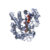 1ry0S S: Starting model for refinement |
|---|---|
| Similar structure data |


-Links
 PDBj
PDBj


- Assembly
| Deposited unit | 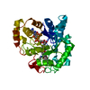
| ||||||||
|---|---|---|---|---|---|---|---|---|---|
| 1 |
| ||||||||
| Unit cell |
| ||||||||
| Details | Monomeric protein containing one NADPH and one bimatoprost. |
-Components
| #1: Protein | Mass: 36896.215 Da / Num. of mol.: 1 Source method: isolated from a genetically manipulated source Source: (gene. exp.) Homo sapiens (human) / Gene: AKR1C3, DDH1, KIAA0119, PGFS / Plasmid: pUC-hLuFS / Production host:  References: UniProt: P42330, trans-1,2-dihydrobenzene-1,2-diol dehydrogenase, EC: 1.1.1.213, prostaglandin-F synthase, 17beta-estradiol 17-dehydrogenase |
|---|---|
| #2: Chemical | ChemComp-NAP /   Mass: 743.405 Da / Num. of mol.: 1 / Source method: obtained synthetically / Formula: C21H28N7O17P3 Mass: 743.405 Da / Num. of mol.: 1 / Source method: obtained synthetically / Formula: C21H28N7O17P3 |
| #3: Chemical | ChemComp-15M / (  Mass: 415.566 Da / Num. of mol.: 1 / Source method: obtained synthetically / Formula: C25H37NO4 Mass: 415.566 Da / Num. of mol.: 1 / Source method: obtained synthetically / Formula: C25H37NO4 |
| #4: Water | ChemComp-HOH /  Mass: 18.015 Da / Num. of mol.: 115 / Source method: isolated from a natural source / Formula: H2O Mass: 18.015 Da / Num. of mol.: 115 / Source method: isolated from a natural source / Formula: H2O |
-Experimental details
-Experiment
| Experiment | Method: X-RAY DIFFRACTION / Number of used crystals: 1 |
|---|
- Sample preparation
| Crystal | Density Matthews: 2.26 Å3/Da / Density % sol: 45.46 % |
|---|---|
| Crystal grow | Temperature: 277 K / Method: vapor diffusion, hanging drop / pH: 7 Details: 1.0 mM BMP, 1.0 mM NADPH, 0.14 M NaCl, 50 mM MES, 26% PEG-8000, pH 7.0, VAPOR DIFFUSION, HANGING DROP, temperature 277K |
-Data collection
| Diffraction | Mean temperature: 95 K |
|---|---|
| Diffraction source | Source: ROTATING ANODE / Type: RIGAKU RU200 / Wavelength: 1.5418 Å |
| Detector | Type: RIGAKU RAXIS IIC / Detector: IMAGE PLATE / Date: May 5, 2005 / Details: confocal optics |
| Radiation | Protocol: SINGLE WAVELENGTH / Monochromatic (M) / Laue (L): M / Scattering type: x-ray |
| Radiation wavelength | Wavelength: 1.5418 Å / Relative weight: 1 |
| Reflection | Resolution: 2→20 Å / Num. all: 19525 / Num. obs: 19525 / % possible obs: 89 % / Observed criterion σ(F): 0 / Observed criterion σ(I): 0 |
| Reflection shell | Resolution: 2→2.1 Å / % possible all: 63 |
- Processing
| Software |
| ||||||||||||||||||||
|---|---|---|---|---|---|---|---|---|---|---|---|---|---|---|---|---|---|---|---|---|---|
| Refinement | Method to determine structure: MOLECULAR REPLACEMENT Starting model: pdb entry 1RY0 Resolution: 2→20 Å / σ(F): 0 / Stereochemistry target values: Engh & Huber
| ||||||||||||||||||||
| Refinement step | Cycle: LAST / Resolution: 2→20 Å
|
