[English] 日本語
Yorodumi
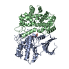
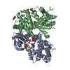
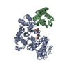
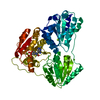
- PDB-2f2c: X-ray structure of human CDK6-Vcyclinwith the inhibitor aminopurv... -

+
Open data


ID or keywords:

Loading...

-
Basic information

Entry
Database: PDB / ID: 2f2c
TitleX-ray structure of human CDK6-Vcyclinwith the inhibitor aminopurvalanol
Components
  • Cell division protein kinase 6
  • Cyclin homolog
KeywordsCELL CYCLE/TRANSFERASE / Small molecule inhibitor bound between N-terminal and C-terminal domain of kinase / CELL CYCLE-TRANSFERASE COMPLEX
Function / homology
Function and homology information


cyclin D2-CDK6 complex / cell dedifferentiation / Drug-mediated inhibition of CDK4/CDK6 activity / Evasion of Oncogene Induced Senescence Due to Defective p16INK4A binding to CDK4 and CDK6 / Evasion of Oxidative Stress Induced Senescence Due to Defective p16INK4A binding to CDK4 and CDK6 / FBXO family protein binding / lateral ventricle development / negative regulation of myeloid cell differentiation / type B pancreatic cell development / negative regulation of monocyte differentiation ...cyclin D2-CDK6 complex / cell dedifferentiation / Drug-mediated inhibition of CDK4/CDK6 activity / Evasion of Oncogene Induced Senescence Due to Defective p16INK4A binding to CDK4 and CDK6 / Evasion of Oxidative Stress Induced Senescence Due to Defective p16INK4A binding to CDK4 and CDK6 / FBXO family protein binding / lateral ventricle development / negative regulation of myeloid cell differentiation / type B pancreatic cell development / negative regulation of monocyte differentiation / astrocyte development / dentate gyrus development / gliogenesis / regulation of cell motility / Regulation of RUNX1 Expression and Activity / regulation of hematopoietic stem cell differentiation / cyclin-dependent protein serine/threonine kinase regulator activity / positive regulation of cell-matrix adhesion / generation of neurons / Defective binding of RB1 mutants to E2F1,(E2F2, E2F3) / negative regulation of cellular senescence / negative regulation of cell differentiation / negative regulation of cell cycle / cyclin-dependent protein kinase holoenzyme complex / hematopoietic stem cell differentiation / negative regulation of osteoblast differentiation / cyclin-dependent kinase / cyclin-dependent protein serine/threonine kinase activity / Notch signaling pathway / ruffle / regulation of G2/M transition of mitotic cell cycle / cyclin binding / : / response to virus / regulation of erythrocyte differentiation / Oncogene Induced Senescence / G1/S transition of mitotic cell cycle / Cyclin D associated events in G1 / negative regulation of epithelial cell proliferation / positive regulation of fibroblast proliferation / T cell differentiation in thymus / Senescence-Associated Secretory Phenotype (SASP) / regulation of gene expression / Oxidative Stress Induced Senescence / regulation of cell cycle / cell cycle / negative regulation of cell population proliferation / cell division / protein phosphorylation / protein serine kinase activity / centrosome / positive regulation of gene expression / negative regulation of transcription by RNA polymerase II / signal transduction / nucleoplasm / ATP binding / nucleus / cytoplasm / cytosol
Similarity search - Function
Cyclin domain, herpesvirus / Herpesviridae viral cyclin / Cyclin, herpesvirus / Cyclin-dependent kinase 6 / : / Cyclins signature. / Cyclin / Cyclin-like / Cyclin A; domain 1 / Cyclin, N-terminal ...Cyclin domain, herpesvirus / Herpesviridae viral cyclin / Cyclin, herpesvirus / Cyclin-dependent kinase 6 / : / Cyclins signature. / Cyclin / Cyclin-like / Cyclin A; domain 1 / Cyclin, N-terminal / Cyclin, N-terminal domain / Cyclin-like / domain present in cyclins, TFIIB and Retinoblastoma / Cyclin-like superfamily / Transferase(Phosphotransferase) domain 1 / Transferase(Phosphotransferase); domain 1 / Phosphorylase Kinase; domain 1 / Phosphorylase Kinase; domain 1 / Serine/threonine-protein kinase, active site / Serine/Threonine protein kinases active-site signature. / Protein kinase domain / Serine/Threonine protein kinases, catalytic domain / Protein kinase, ATP binding site / Protein kinases ATP-binding region signature. / Protein kinase domain profile. / Protein kinase domain / Protein kinase-like domain superfamily / 2-Layer Sandwich / Orthogonal Bundle / Mainly Alpha / Alpha Beta
Similarity search - Domain/homology
Chem-AP9 / Cyclin-dependent kinase 6 / Cyclin homolog
Similarity search - Component
Biological speciesHerpesvirus saimiri
Homo sapiens (human)
MethodX-RAY DIFFRACTION / SYNCHROTRON / MOLECULAR REPLACEMENT / Resolution: 2.8 Å
AuthorsSchulze-Gahmen, U. / Lu, H.
CitationJournal: J.Med.Chem. / Year: 2006
Title: Toward understanding the structural basis of cyclin-dependent kinase 6 specific inhibition.
Authors: Lu, H. / Schulze-Gahmen, U.
History
DepositionNov 16, 2005Deposition site: RCSB / Processing site: RCSB
Revision 1.0Jun 13, 2006Provider: repository / Type: Initial release
Revision 1.1May 1, 2008Group: Version format compliance
Revision 1.2Jul 13, 2011Group: Advisory / Version format compliance
Revision 1.3Jan 24, 2018Group: Structure summary / Category: audit_author / Item: _audit_author.name
Revision 1.4Feb 14, 2024Group: Data collection / Database references / Derived calculations
Category: chem_comp_atom / chem_comp_bond ...chem_comp_atom / chem_comp_bond / database_2 / struct_site
Item: _database_2.pdbx_DOI / _database_2.pdbx_database_accession ..._database_2.pdbx_DOI / _database_2.pdbx_database_accession / _struct_site.pdbx_auth_asym_id / _struct_site.pdbx_auth_comp_id / _struct_site.pdbx_auth_seq_id

-
Structure visualization

Structure viewerMolecule:
MolmilJmol/JSmol

Downloads & links

-
Assembly

Deposited unit
A: Cyclin homolog
B: Cell division protein kinase 6
hetero molecules


Theoretical massNumber of molelcules
Total (without water)64,3105
Polymers63,7322
Non-polymers5783
Water00
1


  • Idetical with deposited unit
  • defined by author&software
TypeNameSymmetry operationNumber
identity operation1_555x,y,z1
Buried area5570 Å2
ΔGint-37 kcal/mol
Surface area22630 Å2
MethodPISA
Unit cell
Length a, b, c (Å)71.517, 71.517, 449.297
Angle α, β, γ (deg.)90.00, 90.00, 120.00
Int Tables number179
Space group name H-MP6522

-
Components

#1: Protein Cyclin homolog / V-cyclin


Mass: 28665.416 Da / Num. of mol.: 1
Source method: isolated from a genetically manipulated source
Source: (gene. exp.) 
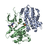
Herpesvirus saimiri (strain 11) / Genus: Rhadinovirus / Species: Saimiriine herpesvirus 2 / Strain: 11 / Gene: 72, ECLF2 / Production host: Spodoptera frugiperda (fall armyworm) / References: UniProt: Q01043
#2: Protein Cell division protein kinase 6 / Serine/threonine-protein kinase PLSTIRE


Mass: 35066.332 Da / Num. of mol.: 1 / Fragment: fragment 1-308
Source method: isolated from a genetically manipulated source
Source: (gene. exp.) Homo sapiens (human) / Gene: CDK6 / Production host: Spodoptera frugiperda (fall armyworm) / References: UniProt: Q00534, EC: 2.7.1.37
#3: Chemical ChemComp-SO4 / SULFATE ION


Mass: 96.063 Da / Num. of mol.: 1 / Source method: obtained synthetically / Formula: SO4
#4: Chemical ChemComp-AP9 / (2S)-2-({6-[(3-AMINO-5-CHLOROPHENYL)AMINO]-9-ISOPROPYL-9H-PURIN-2-YL}AMINO)-3-METHYLBUTAN-1-OL / AMINOPURVALANOL


Mass: 403.909 Da / Num. of mol.: 1 / Source method: obtained synthetically / Formula: C19H26ClN7O
#5: Chemical ChemComp-DMS / DIMETHYL SULFOXIDE


Mass: 78.133 Da / Num. of mol.: 1 / Source method: obtained synthetically / Formula: C2H6OS / Comment: DMSO, precipitant*YM

-
Experimental details

-
Experiment

ExperimentMethod: X-RAY DIFFRACTION / Number of used crystals: 1

-
Sample preparation

CrystalDensity Matthews: 2.6 Å3/Da / Density % sol: 52.71 %
Crystal growTemperature: 293 K / Method: vapor diffusion, hanging drop / pH: 8
Details: 50 mM Tris/HCl pH 8.0, 0.1 M CaOAc, 13% PEG 3350, 10 mM DTT, 0.1M sulfobetaine, 1mM inhibitor, VAPOR DIFFUSION, HANGING DROP, temperature 293K

-
Data collection

DiffractionMean temperature: 100 K
Diffraction sourceSource: SYNCHROTRON / Site: ALS / Beamline: 5.0.1 / Wavelength: 1 Å
DetectorType: ADSC QUANTUM 4 / Detector: CCD
RadiationProtocol: SINGLE WAVELENGTH / Monochromatic (M) / Laue (L): M / Scattering type: x-ray
Radiation wavelengthWavelength: 1 Å / Relative weight: 1
ReflectionResolution: 2.78→50 Å / Num. all: 18422 / Num. obs: 17833 / % possible obs: 96.8 % / Observed criterion σ(F): 0 / Observed criterion σ(I): 0 / Rsym value: 0.09 / Net I/σ(I): 15.4
Reflection shellResolution: 2.78→2.88 Å / % possible all: 0.995

-
Processing

Software
NameVersionClassification
REFMAC5.2.0003refinement
HKL-2000data reduction
CCP4(TRUNCATE)data scaling
AMoREphasing
RefinementMethod to determine structure: MOLECULAR REPLACEMENT / Resolution: 2.8→20 Å / Cor.coef. Fo:Fc: 0.919 / Cor.coef. Fo:Fc free: 0.885 / SU B: 40.424 / SU ML: 0.352 / TLS residual ADP flag: LIKELY RESIDUAL / Cross valid method: THROUGHOUT / ESU R: 2.645 / ESU R Free: 0.43 / Stereochemistry target values: MAXIMUM LIKELIHOOD
RfactorNum. reflection% reflectionSelection details
Rfree0.30144 893 5.1 %RANDOM
Rwork0.23774 ---
obs0.24071 16516 96.91 %-
Solvent computationIon probe radii: 0.8 Å / Shrinkage radii: 0.8 Å / VDW probe radii: 1.2 Å / Solvent model: BABINET MODEL WITH MASK
Displacement parametersBiso mean: 73.703 Å2
Baniso -1Baniso -2Baniso -3
1-1.26 Å20.63 Å20 Å2
2--1.26 Å20 Å2
3----1.89 Å2
Refinement stepCycle: LAST / Resolution: 2.8→20 Å
ProteinNucleic acidLigandSolventTotal
Num. atoms4022 0 37 0 4059
Refine LS restraints
Refine-IDTypeDev idealDev ideal targetNumber
X-RAY DIFFRACTIONr_bond_refined_d0.0110.0224135
X-RAY DIFFRACTIONr_angle_refined_deg1.3661.9735632
X-RAY DIFFRACTIONr_dihedral_angle_1_deg5.9675516
X-RAY DIFFRACTIONr_dihedral_angle_2_deg39.20524.452155
X-RAY DIFFRACTIONr_dihedral_angle_3_deg19.4115689
X-RAY DIFFRACTIONr_dihedral_angle_4_deg23.6861518
X-RAY DIFFRACTIONr_chiral_restr0.0920.2688
X-RAY DIFFRACTIONr_gen_planes_refined0.0050.023001
X-RAY DIFFRACTIONr_nbd_refined0.2420.21952
X-RAY DIFFRACTIONr_nbtor_refined0.310.22869
X-RAY DIFFRACTIONr_xyhbond_nbd_refined0.160.2111
X-RAY DIFFRACTIONr_symmetry_vdw_refined0.3110.231
X-RAY DIFFRACTIONr_symmetry_hbond_refined0.2010.22
X-RAY DIFFRACTIONr_mcbond_it0.421.52671
X-RAY DIFFRACTIONr_mcangle_it0.7524219
X-RAY DIFFRACTIONr_scbond_it1.12631661
X-RAY DIFFRACTIONr_scangle_it1.8154.51413
LS refinement shellResolution: 2.8→2.871 Å / Total num. of bins used: 20
RfactorNum. reflection% reflection
Rfree0.433 75 -
Rwork0.336 1193 -
obs--99.92 %
Refinement TLS params.

Method: refined / Refine-ID: X-RAY DIFFRACTION

IDL112)L122)L132)L222)L232)L332)S11 (Å °)S12 (Å °)S13 (Å °)S21 (Å °)S22 (Å °)S23 (Å °)S31 (Å °)S32 (Å °)S33 (Å °)T112)T122)T132)T222)T232)T332)Origin x (Å)Origin y (Å)Origin z (Å)
14.01830.0785-1.4723.44371.32045.16850.1437-0.39620.0363-0.2811-0.3423-0.3717-0.15740.22110.1985-0.0871-0.2842-0.1111-0.11970.1257-0.215739.01856.92155.436
24.5569-2.0697-0.2835.3557-0.27645.58820.2361-0.3511-1.243-0.3598-0.4861-0.91841.30790.89970.250.4387-0.2244-0.05030.14230.3440.522340.65928.89560.45
33.86672.2213-0.22514.7619-0.98144.1350.1428-0.4321-0.5909-0.2184-0.22420.45350.936-1.10890.08140.1728-0.5464-0.07210.2934-0.00490.02814.70432.99758.29
Refinement TLS group
IDRefine-IDRefine TLS-IDAuth asym-IDLabel asym-IDAuth seq-IDLabel seq-ID
1X-RAY DIFFRACTION1AA8 - 2548 - 254
2X-RAY DIFFRACTION2BB9 - 1009 - 100
3X-RAY DIFFRACTION3BB101 - 305101 - 305
4X-RAY DIFFRACTION3BD401

+
About Yorodumi

-
News

-
Feb 9, 2022. New format data for meta-information of EMDB entries

New format data for meta-information of EMDB entries

  • Version 3 of the EMDB header file is now the official format.
  • The previous official version 1.9 will be removed from the archive.

Related info.:EMDB header

External links:wwPDB to switch to version 3 of the EMDB data model

-
Aug 12, 2020. Covid-19 info

Covid-19 info

URL: https://pdbjlvh1.pdbj.org/emnavi/covid19.php

New page: Covid-19 featured information page in EM Navigator.

Related info.:Covid-19 info / Mar 5, 2020. Novel coronavirus structure data

+
Mar 5, 2020. Novel coronavirus structure data

Novel coronavirus structure data

Related info.:Yorodumi Speices / Aug 12, 2020. Covid-19 info

External links:COVID-19 featured content - PDBj / Molecule of the Month (242):Coronavirus Proteases

+
Jan 31, 2019. EMDB accession codes are about to change! (news from PDBe EMDB page)

EMDB accession codes are about to change! (news from PDBe EMDB page)

  • The allocation of 4 digits for EMDB accession codes will soon come to an end. Whilst these codes will remain in use, new EMDB accession codes will include an additional digit and will expand incrementally as the available range of codes is exhausted. The current 4-digit format prefixed with “EMD-” (i.e. EMD-XXXX) will advance to a 5-digit format (i.e. EMD-XXXXX), and so on. It is currently estimated that the 4-digit codes will be depleted around Spring 2019, at which point the 5-digit format will come into force.
  • The EM Navigator/Yorodumi systems omit the EMD- prefix.

Related info.:Q: What is EMD? / ID/Accession-code notation in Yorodumi/EM Navigator

External links:EMDB Accession Codes are Changing Soon! / Contact to PDBj

+
Jul 12, 2017. Major update of PDB

Major update of PDB

  • wwPDB released updated PDB data conforming to the new PDBx/mmCIF dictionary.
  • This is a major update changing the version number from 4 to 5, and with Remediation, in which all the entries are updated.
  • In this update, many items about electron microscopy experimental information are reorganized (e.g. em_software).
  • Now, EM Navigator and Yorodumi are based on the updated data.

External links:wwPDB Remediation / Enriched Model Files Conforming to OneDep Data Standards Now Available in the PDB FTP Archive

-
Yorodumi

Thousand views of thousand structures

  • Yorodumi is a browser for structure data from EMDB, PDB, SASBDB, etc.
  • This page is also the successor to EM Navigator detail page, and also detail information page/front-end page for Omokage search.
  • The word "yorodu" (or yorozu) is an old Japanese word meaning "ten thousand". "mi" (miru) is to see.

Related info.:EMDB / PDB / SASBDB / Comparison of 3 databanks / Yorodumi Search / Aug 31, 2016. New EM Navigator & Yorodumi / Yorodumi Papers / Jmol/JSmol / Function and homology information / Changes in new EM Navigator and Yorodumi

Read more