 Movie
Movie Controller


+ Open data

- Basic information
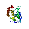
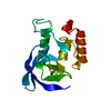
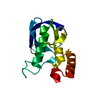
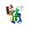
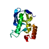
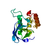
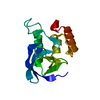
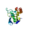
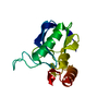
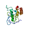
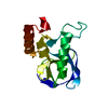
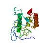
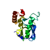
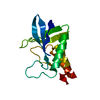
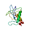
| Entry | Database: PDB / ID: 2f0d | ||||||
|---|---|---|---|---|---|---|---|
| Title | Crystal structure of Staphylococcal nuclease mutant I92V | ||||||
 Components Components | Staphylococcal nuclease | ||||||
 Keywords Keywords | HYDROLASE / DNA HYDROLASE / RNA HYDROLASE / ENDONUCLEASE / CALCIUM / SIGNAL | ||||||
| Function / homology |  Function and homology information Function and homology informationmicrococcal nuclease / 3' overhang single-stranded DNA endonuclease activity / nucleic acid binding / extracellular region / membrane / metal ion binding Similarity search - Function | ||||||
| Biological species |   Staphylococcus aureus (bacteria) Staphylococcus aureus (bacteria) | ||||||
| Method |  X-RAY DIFFRACTION / MOLECULAR REPLACEMENT / Resolution: 2.5 Å X-RAY DIFFRACTION / MOLECULAR REPLACEMENT / Resolution: 2.5 Å | ||||||
 Authors Authors | Lu, J.Z. / Sakon, J. / Stites, W.E. | ||||||
 Citation Citation | Journal: To be Published Title: Hydrophobic core mutants of Staphylococcal nuclease Authors: Lu, J.Z. / Sakon, J. / Stites, W.E. | ||||||
| History |
|
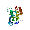
- Structure visualization
| Structure viewer | Molecule: MolmilJmol/JSmol |
|---|
- Downloads & links
-Download
| PDBx/mmCIF format | 2f0d.cif.gz | 38.3 KB | Display | PDBx/mmCIF format |
|---|---|---|---|---|
| PDB format | pdb2f0d.ent.gz | 26.8 KB | Display | PDB format |
| PDBx/mmJSON format | 2f0d.json.gz | Tree view | PDBx/mmJSON format | |
| Others |  Other downloads Other downloads |
-Validation report
| Arichive directory | https://data.pdbj.org/pub/pdb/validation_reports/f0/2f0dftp://data.pdbj.org/pub/pdb/validation_reports/f0/2f0d | HTTPS FTP |
|---|
-Related structure data
| Related structure data | 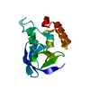 2f0eC 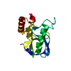 2f0fC 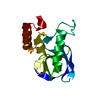 2f0gC 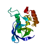 2f0hC  2f0iC  2f0jC  2f0kC  2f0lC 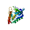 2f0mC 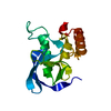 2f0nC 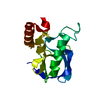 2f0oC  2f0pC  2f0qC 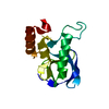 2f0sC 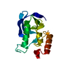 2f0tC 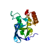 2f0uC 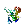 2f0vC  2f0wC  4h7bC  4i65C  4k8iC  4k8jC C: citing same article ( |
|---|---|
| Similar structure data |


-Links
 PDBj
PDBj- Assembly
| Deposited unit | 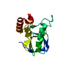
| ||||||||
|---|---|---|---|---|---|---|---|---|---|
| 1 |
| ||||||||
| Unit cell |
|
-Components
| #1: Protein | Mass: 16829.303 Da / Num. of mol.: 1 / Mutation: I92V Source method: isolated from a genetically manipulated source Source: (gene. exp.) Staphylococcus aureus (bacteria) / Gene: nuc / Plasmid: pL13 / Production host: |
|---|---|
| #2: Water | ChemComp-HOH /  Mass: 18.015 Da / Num. of mol.: 12 / Source method: isolated from a natural source / Formula: H2O Mass: 18.015 Da / Num. of mol.: 12 / Source method: isolated from a natural source / Formula: H2O |
-Experimental details
-Experiment
| Experiment | Method: X-RAY DIFFRACTION / Number of used crystals: 1 |
|---|
- Sample preparation
| Crystal | Density Matthews: 2.1 Å3/Da / Density % sol: 44 % |
|---|---|
| Crystal grow | Temperature: 277 K / Method: vapor diffusion, hanging drop / pH: 7.5 Details: 50mM phosphate buffer, 45% MPD, pH 7.5, VAPOR DIFFUSION, HANGING DROP, temperature 277K |
-Data collection
| Diffraction | Mean temperature: 293 K |
|---|---|
| Diffraction source | Source: ROTATING ANODE / Type: RIGAKU / Wavelength: 1.5418 Å |
| Detector | Type: RIGAKU RAXIS IV / Detector: IMAGE PLATE |
| Radiation | Protocol: SINGLE WAVELENGTH / Monochromatic (M) / Laue (L): M / Scattering type: x-ray |
| Radiation wavelength | Wavelength: 1.5418 Å / Relative weight: 1 |
| Reflection | Resolution: 2.5→20 Å / Num. all: 5099 / Num. obs: 4786 / % possible obs: 93.9 % / Observed criterion σ(I): 0 |
- Processing
| Software |
| ||||||||||||||||||||||||||||
|---|---|---|---|---|---|---|---|---|---|---|---|---|---|---|---|---|---|---|---|---|---|---|---|---|---|---|---|---|---|
| Refinement | Method to determine structure: MOLECULAR REPLACEMENT / Resolution: 2.5→20 Å / Data cutoff high absF: 10000 / Data cutoff low absF: 0 / σ(F): 1 / Stereochemistry target values: Engh & Huber
| ||||||||||||||||||||||||||||
| Displacement parameters |
| ||||||||||||||||||||||||||||
| Refinement step | Cycle: LAST / Resolution: 2.5→20 Å
| ||||||||||||||||||||||||||||
| Refine LS restraints |
|
