 Movie
Movie Controller


+ Open data

- Basic information
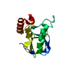
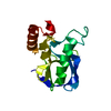
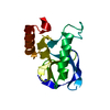
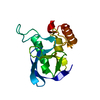
| Entry | Database: PDB / ID: 2eyj | ||||||
|---|---|---|---|---|---|---|---|
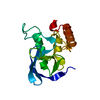
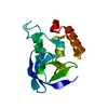
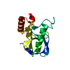
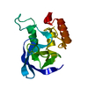
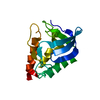
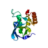
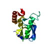
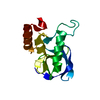
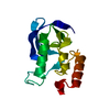
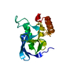
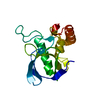
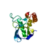
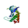
| Title | Crystal structure of Staphylococcal nuclease mutant T62V | ||||||
 Components Components | Staphylococcal nuclease | ||||||
 Keywords Keywords | HYDROLASE / DNA HYDROLASE / RNA HYDROLASE / ENDONUCLEASE / CALCIUM / SIGNAL | ||||||
| Function / homology |  Function and homology information Function and homology informationmicrococcal nuclease / 3' overhang single-stranded DNA endonuclease activity / nucleic acid binding / extracellular region / membrane / metal ion binding Similarity search - Function | ||||||
| Biological species |   Staphylococcus aureus (bacteria) Staphylococcus aureus (bacteria) | ||||||
| Method |  X-RAY DIFFRACTION / MOLECULAR REPLACEMENT / Resolution: 1.85 Å X-RAY DIFFRACTION / MOLECULAR REPLACEMENT / Resolution: 1.85 Å | ||||||
 Authors Authors | Lu, J.Z. / Sakon, J. / Stites, W.E. | ||||||
 Citation Citation | Journal: To be Published Title: Threonine mutants of Staphylococcal nuclease Authors: Lu, J.Z. / Sakon, J. / Stites, W.E. | ||||||
| History |
|
- Structure visualization
| Structure viewer | Molecule: MolmilJmol/JSmol |
|---|
- Downloads & links
-Download
| PDBx/mmCIF format | 2eyj.cif.gz | 41.4 KB | Display | PDBx/mmCIF format |
|---|---|---|---|---|
| PDB format | pdb2eyj.ent.gz | 28.8 KB | Display | PDB format |
| PDBx/mmJSON format | 2eyj.json.gz | Tree view | PDBx/mmJSON format | |
| Others |  Other downloads Other downloads |
-Validation report
| Arichive directory | https://data.pdbj.org/pub/pdb/validation_reports/ey/2eyjftp://data.pdbj.org/pub/pdb/validation_reports/ey/2eyj | HTTPS FTP |
|---|
-Related structure data
| Related structure data | 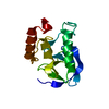 2exzC 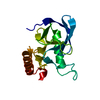 2ey1C  2ey2C 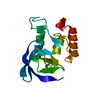 2ey5C 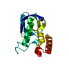 2ey6C  2eyfC 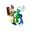 2eyhC 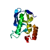 2eylC 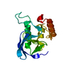 2eymC 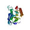 2eyoC  2eypC C: citing same article ( |
|---|---|
| Similar structure data |


-Links
 PDBj
PDBj- Assembly
| Deposited unit | 
| ||||||||
|---|---|---|---|---|---|---|---|---|---|
| 1 |
| ||||||||
| Unit cell |
|
-Components
| #1: Protein | Mass: 16841.355 Da / Num. of mol.: 1 / Mutation: T62V Source method: isolated from a genetically manipulated source Source: (gene. exp.) Staphylococcus aureus (bacteria) / Gene: nuc / Plasmid: pL13 / Production host: |
|---|---|
| #2: Water | ChemComp-HOH /  Mass: 18.015 Da / Num. of mol.: 69 / Source method: isolated from a natural source / Formula: H2O Mass: 18.015 Da / Num. of mol.: 69 / Source method: isolated from a natural source / Formula: H2O |
-Experimental details
-Experiment
| Experiment | Method: X-RAY DIFFRACTION / Number of used crystals: 1 |
|---|
- Sample preparation
| Crystal | Density Matthews: 2.06 Å3/Da / Density % sol: 43.6 % |
|---|---|
| Crystal grow | Temperature: 277 K / Method: vapor diffusion, hanging drop / pH: 7.5 Details: 50mM phosphate buffer, 45% MPD, pH 7.5, VAPOR DIFFUSION, HANGING DROP, temperature 277K |
-Data collection
| Diffraction | Mean temperature: 293 K |
|---|---|
| Diffraction source | Source: ROTATING ANODE / Type: RIGAKU / Wavelength: 1.5418 Å |
| Detector | Type: RIGAKU RAXIS IV / Detector: IMAGE PLATE |
| Radiation | Protocol: SINGLE WAVELENGTH / Monochromatic (M) / Laue (L): M / Scattering type: x-ray |
| Radiation wavelength | Wavelength: 1.5418 Å / Relative weight: 1 |
| Reflection | Resolution: 1.85→8 Å / Num. all: 10960 / Num. obs: 10960 / Observed criterion σ(I): 0 |
- Processing
| Software |
| |||||||||||||||||||||||||||||||||
|---|---|---|---|---|---|---|---|---|---|---|---|---|---|---|---|---|---|---|---|---|---|---|---|---|---|---|---|---|---|---|---|---|---|---|
| Refinement | Method to determine structure: MOLECULAR REPLACEMENT / Resolution: 1.85→6 Å / Num. parameters: 4771 / Num. restraintsaints: 4581 / Cross valid method: FREE R / σ(F): 0 / Stereochemistry target values: ENGH & HUBER
| |||||||||||||||||||||||||||||||||
| Refine analyze | Num. disordered residues: 8 / Occupancy sum hydrogen: 0 / Occupancy sum non hydrogen: 1160 | |||||||||||||||||||||||||||||||||
| Refinement step | Cycle: LAST / Resolution: 1.85→6 Å
| |||||||||||||||||||||||||||||||||
| Refine LS restraints |
|
