 Movie
Movie Controller

[English] 日本語
 Yorodumi
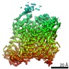
- PDB-2e7p: Crystal structure of the holo form of glutaredoxin C1 from populu... -
+ Open data

- Basic information
| Entry | Database: PDB / ID: 2e7p | ||||||
|---|---|---|---|---|---|---|---|
| Title | Crystal structure of the holo form of glutaredoxin C1 from populus tremula x tremuloides | ||||||
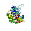
 Components Components | Glutaredoxin | ||||||
 Keywords Keywords | ELECTRON TRANSPORT / THIOREDOXIN FOLD / GLUTAREDOXIN / POPLAR | ||||||
| Function / homology |  Function and homology information Function and homology informationglutathione disulfide oxidoreductase activity / 2 iron, 2 sulfur cluster binding / cellular response to oxidative stress / metal ion binding / identical protein binding / cytoplasm Similarity search - Function | ||||||
| Biological species |  | ||||||
| Method |  X-RAY DIFFRACTION / SYNCHROTRON / MAD / Resolution: 2.1 Å X-RAY DIFFRACTION / SYNCHROTRON / MAD / Resolution: 2.1 Å | ||||||
 Authors Authors | Unno, H. / Takahashi, T. / Kawakami, T. / Aimoto, S. / Hase, T. / Kusunoki, M. / Rouhier, N. / Jacquot, J.P. | ||||||
 Citation Citation | Journal: Proc.Natl.Acad.Sci.Usa / Year: 2007 Title: Functional, structural, and spectroscopic characterization of a glutathione-ligated [2Fe-2S] cluster in poplar glutaredoxin C1 Authors: Rouhier, N. / Unno, H. / Bandyopadhyay, S. / Masip, L. / Kim, S.K. / Hirasawa, M. / Gualberto, J.M. / Lattard, V. / Kusunoki, M. / Knaff, D.B. / Georgiou, G. / Hase, T. / Johnson, M.K. / Jacquot, J.P. | ||||||
| History |
|
- Structure visualization
| Structure viewer | Molecule: MolmilJmol/JSmol |
|---|
- Downloads & links
-Download
| PDBx/mmCIF format | 2e7p.cif.gz | 101.5 KB | Display | PDBx/mmCIF format |
|---|---|---|---|---|
| PDB format | pdb2e7p.ent.gz | 78.9 KB | Display | PDB format |
| PDBx/mmJSON format | 2e7p.json.gz | Tree view | PDBx/mmJSON format | |
| Others |  Other downloads Other downloads |
-Validation report
| Summary document | 2e7p_validation.pdf.gz | 1.4 MB | Display | wwPDB validaton report |
|---|---|---|---|---|
| Full document | 2e7p_full_validation.pdf.gz | 1.4 MB | Display | |
| Data in XML | 2e7p_validation.xml.gz | 21.8 KB | Display | |
| Data in CIF | 2e7p_validation.cif.gz | 31 KB | Display | |
| Arichive directory | https://data.pdbj.org/pub/pdb/validation_reports/e7/2e7pftp://data.pdbj.org/pub/pdb/validation_reports/e7/2e7p | HTTPS FTP |
-Related structure data
| Similar structure data |
|---|


-Links
 PDBj
PDBj


- Assembly
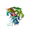
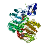
| Deposited unit | 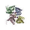
| ||||||||
|---|---|---|---|---|---|---|---|---|---|
| 1 |
| ||||||||
| 2 | 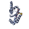
| ||||||||
| 3 | 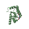
| ||||||||
| 4 | 
| ||||||||
| 5 | 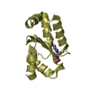
| ||||||||
| Unit cell |
|
-Components
| #1: Protein | Mass: 12435.191 Da / Num. of mol.: 4 / Fragment: RESIDUES 1-116 Source method: isolated from a genetically manipulated source Source: (gene. exp.) Plasmid: PET-3D / Production host:  #2: Chemical | ChemComp-FES / |   Mass: 175.820 Da / Num. of mol.: 1 / Source method: obtained synthetically / Formula: Fe2S2 Mass: 175.820 Da / Num. of mol.: 1 / Source method: obtained synthetically / Formula: Fe2S2#3: Chemical | ChemComp-GSH /   Mass: 307.323 Da / Num. of mol.: 4 / Source method: obtained synthetically / Formula: C10H17N3O6S Mass: 307.323 Da / Num. of mol.: 4 / Source method: obtained synthetically / Formula: C10H17N3O6S#4: Water | ChemComp-HOH / |  Mass: 18.015 Da / Num. of mol.: 293 / Source method: isolated from a natural source / Formula: H2O Mass: 18.015 Da / Num. of mol.: 293 / Source method: isolated from a natural source / Formula: H2O |
|---|
-Experimental details
-Experiment
| Experiment | Method: X-RAY DIFFRACTION / Number of used crystals: 1 |
|---|
- Sample preparation
| Crystal | Density Matthews: 2.54 Å3/Da / Density % sol: 51.53 % |
|---|---|
| Crystal grow | Temperature: 277 K / Method: vapor diffusion, hanging drop / pH: 5 Details: 20% PEG6000, 0.1M CITRIC ACID, pH 5.0, VAPOR DIFFUSION, HANGING DROP, temperature 277K |
-Data collection
| Diffraction |
| |||||||||||||||
|---|---|---|---|---|---|---|---|---|---|---|---|---|---|---|---|---|
| Diffraction source |
| |||||||||||||||
| Detector |
| |||||||||||||||
| Radiation |
| |||||||||||||||
| Radiation wavelength |
| |||||||||||||||
| Reflection | Resolution: 2.1→50 Å / Num. obs: 28968 / % possible obs: 100 % / Redundancy: 11.4 % / Biso Wilson estimate: 31.7 Å2 / Rmerge(I) obs: 0.062 | |||||||||||||||
| Reflection shell | Resolution: 2.1→2.18 Å / Redundancy: 11.4 % / Rmerge(I) obs: 0.345 / % possible all: 100 |

- Processing
| Software |
| ||||||||||||||||||||||||||||||||||||||||||||||||||||||||||||||||||||||||||||||||||||||||||
|---|---|---|---|---|---|---|---|---|---|---|---|---|---|---|---|---|---|---|---|---|---|---|---|---|---|---|---|---|---|---|---|---|---|---|---|---|---|---|---|---|---|---|---|---|---|---|---|---|---|---|---|---|---|---|---|---|---|---|---|---|---|---|---|---|---|---|---|---|---|---|---|---|---|---|---|---|---|---|---|---|---|---|---|---|---|---|---|---|---|---|---|
| Refinement | Method to determine structure: MAD / Resolution: 2.1→24.75 Å / Cor.coef. Fo:Fc: 0.956 / Cor.coef. Fo:Fc free: 0.942 / SU B: 4.558 / SU ML: 0.122 / Cross valid method: THROUGHOUT / ESU R: 0.209 / ESU R Free: 0.172 / Stereochemistry target values: MAXIMUM LIKELIHOOD / Details: HYDROGENS HAVE BEEN ADDED IN THE RIDING POSITIONS
| ||||||||||||||||||||||||||||||||||||||||||||||||||||||||||||||||||||||||||||||||||||||||||
| Solvent computation | Ion probe radii: 0.8 Å / Shrinkage radii: 0.8 Å / VDW probe radii: 1.2 Å / Solvent model: MASK | ||||||||||||||||||||||||||||||||||||||||||||||||||||||||||||||||||||||||||||||||||||||||||
| Displacement parameters | Biso mean: 34.617 Å2
| ||||||||||||||||||||||||||||||||||||||||||||||||||||||||||||||||||||||||||||||||||||||||||
| Refinement step | Cycle: LAST / Resolution: 2.1→24.75 Å
| ||||||||||||||||||||||||||||||||||||||||||||||||||||||||||||||||||||||||||||||||||||||||||
| Refine LS restraints |
| ||||||||||||||||||||||||||||||||||||||||||||||||||||||||||||||||||||||||||||||||||||||||||
| LS refinement shell | Resolution: 2.102→2.157 Å / Total num. of bins used: 20
|
