 Movie
Movie Controller

[English] 日本語
 Yorodumi
- PDB-2e7f: 5-methyltetrahydrofolate corrinoid/iron sulfur protein methyltran... -
+ Open data

- Basic information
| Entry | Database: PDB / ID: 2e7f | ||||||
|---|---|---|---|---|---|---|---|
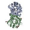
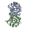
| Title | 5-methyltetrahydrofolate corrinoid/iron sulfur protein methyltransferase complexed with methyltetrahydrofolate to 2.2 Angsrom resolution | ||||||
 Components Components | 5-methyltetrahydrofolate corrinoid/iron sulfur protein methyltransferase | ||||||
 Keywords Keywords | TRANSFERASE / Methyltetrahydrofolate-protein complex / corrionoid / vitamin B12 / TIM Barrel | ||||||
| Function / homology |  Function and homology information Function and homology information5-methyltetrahydrofolate-corrinoid/iron-sulfur protein Co-methyltransferase / methyltetrahydrofolate:corrinoid/iron-sulfur protein methyltransferase activity / methionine synthase activity / homocysteine metabolic process / cobalamin binding / tetrahydrofolate metabolic process / carbon fixation / methyltransferase activity / methylation / calcium ion binding / cytosol Similarity search - Function | ||||||
| Biological species |  Moorella thermoacetica (bacteria) Moorella thermoacetica (bacteria) | ||||||
| Method |  X-RAY DIFFRACTION / SYNCHROTRON / MOLECULAR REPLACEMENT / Resolution: 2.2 Å X-RAY DIFFRACTION / SYNCHROTRON / MOLECULAR REPLACEMENT / Resolution: 2.2 Å | ||||||
 Authors Authors | Doukov, T.I. / Drennan, C.L. / Hemmi, H. / Ragsdale, S.W. | ||||||
 Citation Citation | Journal: J.Biol.Chem. / Year: 2007 Title: Structural and kinetic evidence for an extended hydrogen-bonding network in catalysis of methyl group transfer. Role of an active site asparagine residue in activation of methyl transfer by methyltransferases. Authors: Doukov, T.I. / Hemmi, H. / Drennan, C.L. / Ragsdale, S.W. #1: Journal: Structure / Year: 2000Title: Crystal structure of a methyltetrahydrofolate- and corrinoid-dependent methyltransferase Authors: Doukov, T.I. / Seravalli, J. / Stezowski, J.J. / Ragsdale, S.W. | ||||||
| History |
|
- Structure visualization
| Structure viewer | Molecule: MolmilJmol/JSmol |
|---|
- Downloads & links
-Download
| PDBx/mmCIF format | 2e7f.cif.gz | 123.7 KB | Display | PDBx/mmCIF format |
|---|---|---|---|---|
| PDB format | pdb2e7f.ent.gz | 94.9 KB | Display | PDB format |
| PDBx/mmJSON format | 2e7f.json.gz | Tree view | PDBx/mmJSON format | |
| Others |  Other downloads Other downloads |
-Validation report
| Arichive directory | https://data.pdbj.org/pub/pdb/validation_reports/e7/2e7fftp://data.pdbj.org/pub/pdb/validation_reports/e7/2e7f | HTTPS FTP |
|---|
-Related structure data
| Related structure data |  2ogyC  1f6yS S: Starting model for refinement C: citing same article ( |
|---|---|
| Similar structure data |


-Links
 PDBj
PDBj- Assembly
| Deposited unit | 
| ||||||||
|---|---|---|---|---|---|---|---|---|---|
| 1 |
| ||||||||
| Unit cell |
| ||||||||
| Details | THIS ENTRY CONTAINS THE CRYSTALLOGRAPHIC ASYMMETRIC UNIT WHICH CONSISTS OF 2 CHAIN(S) |
-Components
| #1: Protein | Mass: 28638.119 Da / Num. of mol.: 2 Source method: isolated from a genetically manipulated source Source: (gene. exp.) Moorella thermoacetica (bacteria) / Gene: MeTr, acsE / Plasmid: pET3a / Production host: #2: Chemical |   Mass: 40.078 Da / Num. of mol.: 2 / Source method: obtained synthetically / Formula: Ca Mass: 40.078 Da / Num. of mol.: 2 / Source method: obtained synthetically / Formula: Ca#3: Chemical |   Mass: 459.456 Da / Num. of mol.: 2 / Source method: obtained synthetically / Formula: C20H25N7O6 Mass: 459.456 Da / Num. of mol.: 2 / Source method: obtained synthetically / Formula: C20H25N7O6#4: Water | ChemComp-HOH / |  Mass: 18.015 Da / Num. of mol.: 435 / Source method: isolated from a natural source / Formula: H2O Mass: 18.015 Da / Num. of mol.: 435 / Source method: isolated from a natural source / Formula: H2O |
|---|
-Experimental details
-Experiment
| Experiment | Method: X-RAY DIFFRACTION / Number of used crystals: 1 |
|---|
- Sample preparation
| Crystal | Density Matthews: 2.33 Å3/Da / Density % sol: 47.24 % |
|---|---|
| Crystal grow | Temperature: 298 K / Method: vapor diffusion, hanging drop / pH: 7.5 Details: 8-15% PEGmme 5000, 0.05M Calcium acetate, 20% Glycerol, 0.05M HEPES buffer; 3-fold molar excess of the MTHF substrate (Schricks Labolatories, Jona, Switzerland). The supersaturation of the ...Details: 8-15% PEGmme 5000, 0.05M Calcium acetate, 20% Glycerol, 0.05M HEPES buffer; 3-fold molar excess of the MTHF substrate (Schricks Labolatories, Jona, Switzerland). The supersaturation of the precipitant solution required dilution of the protein-CH3-H4folate complex by 50-100-fold in order to obtain single crystals., pH 7.5, VAPOR DIFFUSION, HANGING DROP, temperature 298K |
-Data collection
| Diffraction | Mean temperature: 100 K |
|---|---|
| Diffraction source | Source: SYNCHROTRON / Site: NSLS  / Beamline: X12C / Wavelength: 0.91938 Å / Beamline: X12C / Wavelength: 0.91938 Å |
| Detector | Type: BRANDEIS - B4 / Detector: CCD / Date: Jul 30, 2000 |
| Radiation | Monochromator: SI(111) or SI(220) / Protocol: SINGLE WAVELENGTH / Monochromatic (M) / Laue (L): M / Scattering type: x-ray |
| Radiation wavelength | Wavelength: 0.91938 Å / Relative weight: 1 |
| Reflection | Resolution: 2.2→30.92 Å / Num. obs: 26806 / % possible obs: 96.2 % / Redundancy: 3.8 % / Biso Wilson estimate: 14.8 Å2 / Rmerge(I) obs: 0.086 / Rsym value: 0.086 / Net I/σ(I): 11.5 |
| Reflection shell | Resolution: 2.2→2.26 Å / Redundancy: 3.5 % / Mean I/σ(I) obs: 3.5 / Num. unique all: 1750 / Rsym value: 0.206 / % possible all: 87.9 |
- Processing
| Software |
| |||||||||||||||||||||||||
|---|---|---|---|---|---|---|---|---|---|---|---|---|---|---|---|---|---|---|---|---|---|---|---|---|---|---|
| Refinement | Method to determine structure: MOLECULAR REPLACEMENT Starting model: PDB ENTRY 1F6Y Resolution: 2.2→30.88 Å / Cross valid method: THROUGHOUT / σ(F): 0 / Stereochemistry target values: MAXIMUM LIKELIHOOD
| |||||||||||||||||||||||||
| Refinement step | Cycle: LAST / Resolution: 2.2→30.88 Å
| |||||||||||||||||||||||||
| Refine LS restraints |
| |||||||||||||||||||||||||
| LS refinement shell | Resolution: 2.2→2.26 Å
|
