 Movie
Movie Controller

[English] 日本語
 Yorodumi
- PDB-2e5f: Crystal Structure of the PH0510 protein from Pyrococcus horikoshi... -
+ Open data

- Basic information
| Entry | Database: PDB / ID: 2e5f | ||||||
|---|---|---|---|---|---|---|---|
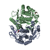
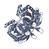
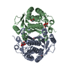
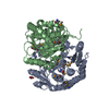
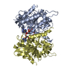
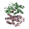
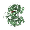
| Title | Crystal Structure of the PH0510 protein from Pyrococcus horikoshii OT3 in complex with phosphate ion | ||||||
 Components Components | Hypothetical protein PH0510 | ||||||
 Keywords Keywords | Structural Genomics / Unknown Function / sugar binding protein / NPPSFA / National Project on Protein Structural and Functional Analyses / RIKEN Structural Genomics/Proteomics Initiative / RSGI | ||||||
| Function / homology |  Function and homology information Function and homology informationcarbohydrate derivative metabolic process / carbohydrate derivative binding Similarity search - Function | ||||||
| Biological species |   Pyrococcus horikoshii (archaea) Pyrococcus horikoshii (archaea) | ||||||
| Method |  X-RAY DIFFRACTION / SYNCHROTRON / MOLECULAR REPLACEMENT / Resolution: 1.35 Å X-RAY DIFFRACTION / SYNCHROTRON / MOLECULAR REPLACEMENT / Resolution: 1.35 Å | ||||||
 Authors Authors | Mizutani, H. / Kunishima, N. / RIKEN Structural Genomics/Proteomics Initiative (RSGI) | ||||||
 Citation Citation | Journal: To be Published Title: Crystal Structure of the PH0510 protein from Pyrococcus horikoshii OT3 Authors: Mizutani, H. / Kunishima, N. | ||||||
| History |
|
- Structure visualization
| Structure viewer | Molecule: MolmilJmol/JSmol |
|---|
- Downloads & links
-Download
| PDBx/mmCIF format | 2e5f.cif.gz | 160.7 KB | Display | PDBx/mmCIF format |
|---|---|---|---|---|
| PDB format | pdb2e5f.ent.gz | 124.3 KB | Display | PDB format |
| PDBx/mmJSON format | 2e5f.json.gz | Tree view | PDBx/mmJSON format | |
| Others |  Other downloads Other downloads |
-Validation report
| Summary document | 2e5f_validation.pdf.gz | 457.8 KB | Display | wwPDB validaton report |
|---|---|---|---|---|
| Full document | 2e5f_full_validation.pdf.gz | 464.3 KB | Display | |
| Data in XML | 2e5f_validation.xml.gz | 33.8 KB | Display | |
| Data in CIF | 2e5f_validation.cif.gz | 53.1 KB | Display | |
| Arichive directory | https://data.pdbj.org/pub/pdb/validation_reports/e5/2e5fftp://data.pdbj.org/pub/pdb/validation_reports/e5/2e5f | HTTPS FTP |
-Related structure data
| Related structure data |  2decSC  2df8C S: Starting model for refinement C: citing same article ( |
|---|---|
| Similar structure data | |
| Other databases |


-Links
 PDBj
PDBj- Assembly
| Deposited unit | 
| ||||||||
|---|---|---|---|---|---|---|---|---|---|
| 1 |
| ||||||||
| Unit cell |
|
-Components
| #1: Protein | Mass: 36855.723 Da / Num. of mol.: 2 Source method: isolated from a genetically manipulated source Source: (gene. exp.) Pyrococcus horikoshii (archaea) / Strain: OT3 / Plasmid: pET11a / Production host:  #2: Chemical |   Mass: 94.971 Da / Num. of mol.: 2 / Source method: obtained synthetically / Formula: PO4 Mass: 94.971 Da / Num. of mol.: 2 / Source method: obtained synthetically / Formula: PO4#3: Chemical | ChemComp-EDO /   Mass: 62.068 Da / Num. of mol.: 5 / Source method: obtained synthetically / Formula: C2H6O2 Mass: 62.068 Da / Num. of mol.: 5 / Source method: obtained synthetically / Formula: C2H6O2#4: Water | ChemComp-HOH / |  Mass: 18.015 Da / Num. of mol.: 886 / Source method: isolated from a natural source / Formula: H2O Mass: 18.015 Da / Num. of mol.: 886 / Source method: isolated from a natural source / Formula: H2O |
|---|
-Experimental details
-Experiment
| Experiment | Method: X-RAY DIFFRACTION / Number of used crystals: 1 |
|---|
- Sample preparation
| Crystal | Density Matthews: 2.14 Å3/Da / Density % sol: 42.4 % |
|---|---|
| Crystal grow | Temperature: 295 K / Method: microbatch / pH: 7.9 Details: 20%(w/v) PEG 3350, 0.2M diammonium hydrogen phosphate, pH 7.9, microbatch, temperature 295K |
-Data collection
| Diffraction | Mean temperature: 100 K |
|---|---|
| Diffraction source | Source: SYNCHROTRON / Site: SPring-8  / Beamline: BL26B1 / Wavelength: 0.8 Å / Beamline: BL26B1 / Wavelength: 0.8 Å |
| Detector | Type: RIGAKU JUPITER 210 / Detector: CCD / Date: Dec 16, 2005 |
| Radiation | Monochromator: bending magnet / Protocol: SINGLE WAVELENGTH / Monochromatic (M) / Laue (L): M / Scattering type: x-ray |
| Radiation wavelength | Wavelength: 0.8 Å / Relative weight: 1 |
| Reflection | Resolution: 1.35→30 Å / Num. all: 135393 / Num. obs: 135393 / % possible obs: 99.7 % / Observed criterion σ(F): 0 / Observed criterion σ(I): 0 / Redundancy: 5.9 % / Biso Wilson estimate: 11.2 Å2 / Rmerge(I) obs: 0.076 / Rsym value: 0.072 / Net I/σ(I): 10.6 |
| Reflection shell | Resolution: 1.35→1.4 Å / Redundancy: 4.4 % / Rmerge(I) obs: 0.276 / Mean I/σ(I) obs: 3.12 / Num. unique all: 13320 / Rsym value: 0.247 / % possible all: 99 |
- Processing
| Software |
| ||||||||||||||||||||||||||||||||||||||||||||||||||||||||||||||||||||||||||||||||
|---|---|---|---|---|---|---|---|---|---|---|---|---|---|---|---|---|---|---|---|---|---|---|---|---|---|---|---|---|---|---|---|---|---|---|---|---|---|---|---|---|---|---|---|---|---|---|---|---|---|---|---|---|---|---|---|---|---|---|---|---|---|---|---|---|---|---|---|---|---|---|---|---|---|---|---|---|---|---|---|---|---|
| Refinement | Method to determine structure: MOLECULAR REPLACEMENT Starting model: PDB ENTRY 2DEC Resolution: 1.35→30 Å / Rfactor Rfree error: 0.002 / Data cutoff high absF: 1430821.6 / Data cutoff low absF: 0 / Isotropic thermal model: RESTRAINED / Cross valid method: THROUGHOUT / σ(F): 0 / Stereochemistry target values: Engh & Huber
| ||||||||||||||||||||||||||||||||||||||||||||||||||||||||||||||||||||||||||||||||
| Solvent computation | Solvent model: FLAT MODEL / Bsol: 51.4136 Å2 / ksol: 0.327727 e/Å3 | ||||||||||||||||||||||||||||||||||||||||||||||||||||||||||||||||||||||||||||||||
| Displacement parameters | Biso mean: 13.7 Å2
| ||||||||||||||||||||||||||||||||||||||||||||||||||||||||||||||||||||||||||||||||
| Refine analyze |
| ||||||||||||||||||||||||||||||||||||||||||||||||||||||||||||||||||||||||||||||||
| Refinement step | Cycle: LAST / Resolution: 1.35→30 Å
| ||||||||||||||||||||||||||||||||||||||||||||||||||||||||||||||||||||||||||||||||
| Refine LS restraints |
| ||||||||||||||||||||||||||||||||||||||||||||||||||||||||||||||||||||||||||||||||
| LS refinement shell | Resolution: 1.35→1.43 Å / Rfactor Rfree error: 0.006 / Total num. of bins used: 6
| ||||||||||||||||||||||||||||||||||||||||||||||||||||||||||||||||||||||||||||||||
| Xplor file |
|
