 Movie
Movie Controller

[English] 日本語
 Yorodumi
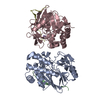
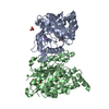
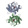
- PDB-2e3j: The crystal structure of epoxide hydrolase B (Rv1938) from mycoba... -
+ Open data

- Basic information
| Entry | Database: PDB / ID: 2e3j | ||||||
|---|---|---|---|---|---|---|---|
| Title | The crystal structure of epoxide hydrolase B (Rv1938) from mycobacterium tuberculosis at 2.1 angstrom | ||||||
 Components Components | EPOXIDE HYDROLASE EPHB | ||||||
 Keywords Keywords | HYDROLASE / EPOXIDE HYDROLASE B / MYCOBACTERIUM TUBERCULOSIS / Structural Genomics / Mycobacterium Tuberculosis Structural Proteomics Project / XMTB | ||||||
| Function / homology |  Function and homology information Function and homology informationsoluble epoxide hydrolase / epoxide hydrolase activity / response to toxic substance / protein homodimerization activity Similarity search - Function | ||||||
| Biological species |   Mycobacterium tuberculosis (bacteria) Mycobacterium tuberculosis (bacteria) | ||||||
| Method |  X-RAY DIFFRACTION / SYNCHROTRON / MOLECULAR REPLACEMENT / Resolution: 2.1 Å X-RAY DIFFRACTION / SYNCHROTRON / MOLECULAR REPLACEMENT / Resolution: 2.1 Å | ||||||
 Authors Authors | Biswal, B.K. / Mycobacterium Tuberculosis Structural Proteomics Project (XMTB) | ||||||
 Citation Citation | Journal: J.Mol.Biol. / Year: 2008 Title: The molecular structure of epoxide hydrolase B from Mycobacterium tuberculosis and its complex with a urea-based inhibitor. Authors: Biswal, B.K. / Morisseau, C. / Garen, G. / Cherney, M.M. / Garen, C. / Niu, C. / Hammock, B.D. / James, M.N. | ||||||
| History |
|
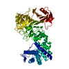
- Structure visualization
| Structure viewer | Molecule: MolmilJmol/JSmol |
|---|
- Downloads & links
-Download
| PDBx/mmCIF format | 2e3j.cif.gz | 84.7 KB | Display | PDBx/mmCIF format |
|---|---|---|---|---|
| PDB format | pdb2e3j.ent.gz | 62.8 KB | Display | PDB format |
| PDBx/mmJSON format | 2e3j.json.gz | Tree view | PDBx/mmJSON format | |
| Others |  Other downloads Other downloads |
-Validation report
| Arichive directory | https://data.pdbj.org/pub/pdb/validation_reports/e3/2e3jftp://data.pdbj.org/pub/pdb/validation_reports/e3/2e3j | HTTPS FTP |
|---|
-Related structure data
| Related structure data | 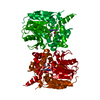 2zjfC 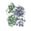 1ek1S S: Starting model for refinement C: citing same article ( |
|---|---|
| Similar structure data | |
| Other databases |


-Links
 PDBj
PDBj


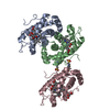
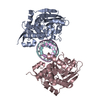
- Assembly
| Deposited unit | 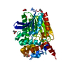
| ||||||||
|---|---|---|---|---|---|---|---|---|---|
| 1 | 
| ||||||||
| 2 |
| ||||||||
| Unit cell |
| ||||||||
| Components on special symmetry positions |
|
-Components
| #1: Protein | Mass: 39305.125 Da / Num. of mol.: 1 Source method: isolated from a genetically manipulated source Source: (gene. exp.) Mycobacterium tuberculosis (bacteria) / Strain: H37Rv / Plasmid: PDEST17 / Production host: | ||
|---|---|---|---|
| #2: Chemical | ChemComp-ACT /   Mass: 59.044 Da / Num. of mol.: 9 / Source method: obtained synthetically / Formula: C2H3O2 Mass: 59.044 Da / Num. of mol.: 9 / Source method: obtained synthetically / Formula: C2H3O2#3: Water | ChemComp-HOH / |  Mass: 18.015 Da / Num. of mol.: 128 / Source method: isolated from a natural source / Formula: H2O Mass: 18.015 Da / Num. of mol.: 128 / Source method: isolated from a natural source / Formula: H2O |
-Experimental details
-Experiment
| Experiment | Method: X-RAY DIFFRACTION / Number of used crystals: 1 |
|---|
- Sample preparation
| Crystal | Density Matthews: 2.19 Å3/Da / Density % sol: 43.89 % |
|---|---|
| Crystal grow | Temperature: 295 K / Method: vapor diffusion, hanging drop / pH: 4.5 Details: 20% 2-PROPANOL, 0.2M CaCl2, 0.1M sodium acetate buffer, protein concentration 5-10mg/ml, pH 4.5, VAPOR DIFFUSION, HANGING DROP, temperature 295K |
-Data collection
| Diffraction | Mean temperature: 100 K |
|---|---|
| Diffraction source | Source: SYNCHROTRON / Site: ALS  / Beamline: 8.3.1 / Wavelength: 1.115869 Å / Beamline: 8.3.1 / Wavelength: 1.115869 Å |
| Detector | Type: ADSC QUANTUM 210 / Detector: CCD / Date: Sep 24, 2005 |
| Radiation | Protocol: SINGLE WAVELENGTH / Monochromatic (M) / Laue (L): M / Scattering type: x-ray |
| Radiation wavelength | Wavelength: 1.115869 Å / Relative weight: 1 |
| Reflection | Resolution: 2.1→39.28 Å / Num. all: 21248 / Num. obs: 20701 / % possible obs: 97.4 % / Observed criterion σ(F): 0 / Observed criterion σ(I): 0 / Redundancy: 6.5 % / Biso Wilson estimate: 35.8 Å2 / Rsym value: 0.07 |
| Reflection shell | Resolution: 2.1→2.18 Å / Redundancy: 3.3 % / Mean I/σ(I) obs: 2.1 / Num. unique all: 1663 / Rsym value: 0.492 / % possible all: 80.4 |
- Processing
| Software |
| |||||||||||||||||||||||||
|---|---|---|---|---|---|---|---|---|---|---|---|---|---|---|---|---|---|---|---|---|---|---|---|---|---|---|
| Refinement | Method to determine structure: MOLECULAR REPLACEMENT Starting model: RESIDUES 245-541 (C-TREMINAL DOMAIN) of PDB ID 1EK1 Resolution: 2.1→39.28 Å / Isotropic thermal model: ISOTROPIC / Cross valid method: THROUGHT / σ(F): 0 / Stereochemistry target values: Engh & Huber
| |||||||||||||||||||||||||
| Displacement parameters | Biso mean: 52.65 Å2
| |||||||||||||||||||||||||
| Refine analyze |
| |||||||||||||||||||||||||
| Refinement step | Cycle: LAST / Resolution: 2.1→39.28 Å
| |||||||||||||||||||||||||
| Refine LS restraints |
| |||||||||||||||||||||||||
| LS refinement shell | Resolution: 2.1→2.23 Å / Rfactor Rfree error: 0.032
|
