[English] 日本語
Yorodumi
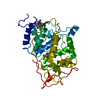
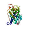
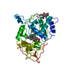
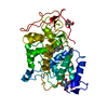
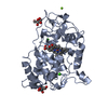
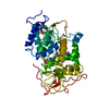
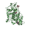
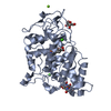
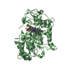
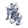
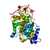
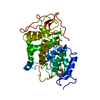
- PDB-2e39: Crystal structure of the CN-bound form of Arthromyces ramosus per... -

+
Open data


ID or keywords:

Loading...

-
Basic information

Entry
Database: PDB / ID: 2.0E+39
TitleCrystal structure of the CN-bound form of Arthromyces ramosus peroxidase at 1.3 Angstroms resolution
ComponentsPeroxidase
KeywordsOXIDOREDUCTASE / heme protein / coordination geometry of heme iron / ARP / peroxidase
Function / homology
Function and homology information


lactoperoxidase activity / peroxidase / hydrogen peroxide catabolic process / cellular response to oxidative stress / heme binding / extracellular region / metal ion binding
Similarity search - Function
Fungal ligninase / Fungal ligninase, C-terminal / Fungal peroxidase extension region / Heme-binding peroxidase Ccp1-like / Peroxidase; domain 2 / Peroxidase, domain 2 / Peroxidase; domain 1 - #10 / Peroxidase; domain 1 / Peroxidases heam-ligand binding site / Peroxidase, active site ...Fungal ligninase / Fungal ligninase, C-terminal / Fungal peroxidase extension region / Heme-binding peroxidase Ccp1-like / Peroxidase; domain 2 / Peroxidase, domain 2 / Peroxidase; domain 1 - #10 / Peroxidase; domain 1 / Peroxidases heam-ligand binding site / Peroxidase, active site / Peroxidases active site signature. / Plant heme peroxidase family profile. / Haem peroxidase / Peroxidase / Peroxidases proximal heme-ligand signature. / Haem peroxidase superfamily / Orthogonal Bundle / Mainly Alpha
Similarity search - Domain/homology
CYANIDE ION / PROTOPORPHYRIN IX CONTAINING FE / alpha-D-mannopyranose / Peroxidase
Similarity search - Component
Biological species'Arthromyces ramosus' (fungus)
MethodX-RAY DIFFRACTION / SYNCHROTRON / FOURIER SYNTHESIS / Resolution: 1.3 Å
AuthorsFukuyama, K. / Okada, T.
Citation
Journal: ACTA CRYSTALLOGR.,SECT.D / Year: 2007
Title: Structures of cyanide, nitric oxide and hydroxylamine complexes of Arthromyces ramosusperoxidase at 100 K refined to 1.3 A resolution: coordination geometries of the ligands to the haem iron
Authors: Fukuyama, K. / Okada, T.
#1: Journal: J.Mol.Biol. / Year: 1994
Title: Crystal structure of the fungal peroxidase from Arthromyces ramosus at 1.9 A resolution. Structural comparisons with the lignin and cytochrome c peroxidases
Authors: Kunishima, N. / Fukuyama, K. / Matsubara, H. / Hatanaka, H. / Shibano, Y. / Amachi, T.
History
DepositionNov 22, 2006Deposition site: PDBJ / Processing site: PDBJ
Revision 1.0Mar 20, 2007Provider: repository / Type: Initial release
Revision 1.1Apr 30, 2008Group: Version format compliance
Revision 1.2Jul 13, 2011Group: Non-polymer description / Version format compliance
Revision 1.3Oct 11, 2017Group: Refinement description / Category: software / Item: _software.classification / _software.name
Revision 2.0Jul 29, 2020Group: Atomic model / Data collection ...Atomic model / Data collection / Derived calculations / Structure summary
Category: atom_site / atom_site_anisotrop ...atom_site / atom_site_anisotrop / chem_comp / entity / pdbx_branch_scheme / pdbx_chem_comp_identifier / pdbx_entity_branch / pdbx_entity_branch_descriptor / pdbx_entity_branch_link / pdbx_entity_branch_list / pdbx_entity_nonpoly / pdbx_nonpoly_scheme / pdbx_struct_assembly_gen / pdbx_struct_conn_angle / struct_asym / struct_conn / struct_conn_type / struct_site / struct_site_gen
Item: _atom_site.auth_asym_id / _atom_site.auth_seq_id ..._atom_site.auth_asym_id / _atom_site.auth_seq_id / _atom_site.label_asym_id / _atom_site_anisotrop.pdbx_auth_asym_id / _atom_site_anisotrop.pdbx_auth_seq_id / _atom_site_anisotrop.pdbx_label_asym_id / _chem_comp.name / _chem_comp.type / _entity.formula_weight / _entity.pdbx_description / _entity.pdbx_number_of_molecules / _entity.type / _pdbx_struct_assembly_gen.asym_id_list / _pdbx_struct_conn_angle.ptnr1_auth_comp_id / _pdbx_struct_conn_angle.ptnr1_auth_seq_id / _pdbx_struct_conn_angle.ptnr1_label_asym_id / _pdbx_struct_conn_angle.ptnr1_label_atom_id / _pdbx_struct_conn_angle.ptnr1_label_comp_id / _pdbx_struct_conn_angle.ptnr1_label_seq_id / _pdbx_struct_conn_angle.ptnr2_auth_comp_id / _pdbx_struct_conn_angle.ptnr2_auth_seq_id / _pdbx_struct_conn_angle.ptnr2_label_asym_id / _pdbx_struct_conn_angle.ptnr2_label_atom_id / _pdbx_struct_conn_angle.ptnr2_label_comp_id / _pdbx_struct_conn_angle.ptnr3_auth_comp_id / _pdbx_struct_conn_angle.ptnr3_auth_seq_id / _pdbx_struct_conn_angle.ptnr3_label_asym_id / _pdbx_struct_conn_angle.ptnr3_label_atom_id / _pdbx_struct_conn_angle.ptnr3_label_comp_id / _pdbx_struct_conn_angle.ptnr3_label_seq_id / _pdbx_struct_conn_angle.value / _struct_conn.conn_type_id / _struct_conn.id / _struct_conn.pdbx_dist_value / _struct_conn.pdbx_leaving_atom_flag / _struct_conn.pdbx_role / _struct_conn.ptnr1_auth_asym_id / _struct_conn.ptnr1_auth_comp_id / _struct_conn.ptnr1_auth_seq_id / _struct_conn.ptnr1_label_asym_id / _struct_conn.ptnr1_label_atom_id / _struct_conn.ptnr1_label_comp_id / _struct_conn.ptnr1_label_seq_id / _struct_conn.ptnr2_auth_asym_id / _struct_conn.ptnr2_auth_comp_id / _struct_conn.ptnr2_auth_seq_id / _struct_conn.ptnr2_label_asym_id / _struct_conn.ptnr2_label_atom_id / _struct_conn.ptnr2_label_comp_id / _struct_conn.ptnr2_label_seq_id / _struct_conn_type.id
Description: Carbohydrate remediation / Provider: repository / Type: Remediation

-
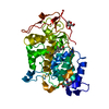
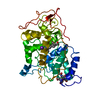
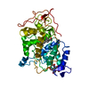
Structure visualization

Structure viewerMolecule:
MolmilJmol/JSmol

Downloads & links

-
Assembly

Deposited unit
A: Peroxidase
hetero molecules


Theoretical massNumber of molelcules
Total (without water)37,0507
Polymers35,7231
Non-polymers1,3276
Water6,972387
1


  • Idetical with deposited unit
  • defined by author
TypeNameSymmetry operationNumber
identity operation1_555x,y,z1
Unit cell
Length a, b, c (Å)73.840, 73.840, 115.680
Angle α, β, γ (deg.)90.00, 90.00, 90.00
Int Tables number94
Space group name H-MP42212
DetailsThe biological assembly is a monomer

-
Components

-
Protein , 1 types, 1 molecules A

#1: Protein Peroxidase /


Mass: 35722.797 Da / Num. of mol.: 1 / Source method: isolated from a natural source / Source: (natural) 'Arthromyces ramosus' (fungus) / Genus: 'Arthromyces' / References: UniProt: P28313, peroxidase

-
Sugars , 2 types, 2 molecules

#2: Polysaccharide 2-acetamido-2-deoxy-beta-D-glucopyranose-(1-4)-2-acetamido-2-deoxy-beta-D-glucopyranose


Type: oligosaccharide / Mass: 424.401 Da / Num. of mol.: 1
Source method: isolated from a genetically manipulated source
DescriptorTypeProgram
DGlcpNAcb1-4DGlcpNAcb1-Glycam Condensed SequenceGMML 1.0
WURCS=2.0/1,2,1/[a2122h-1b_1-5_2*NCC/3=O]/1-1/a4-b1WURCSPDB2Glycan 1.1.0
[]{[(4+1)][b-D-GlcpNAc]{[(4+1)][b-D-GlcpNAc]{}}}LINUCSPDB-CARE
#3: Sugar ChemComp-MAN / alpha-D-mannopyranose / Mannose


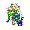
Type: D-saccharide, alpha linking / Mass: 180.156 Da / Num. of mol.: 1
Source method: isolated from a genetically manipulated source
Formula: C6H12O6
IdentifierTypeProgram
DManpaCONDENSED IUPAC CARBOHYDRATE SYMBOLGMML 1.0
a-D-mannopyranoseCOMMON NAMEGMML 1.0
a-D-ManpIUPAC CARBOHYDRATE SYMBOLPDB-CARE 1.0
ManSNFG CARBOHYDRATE SYMBOLGMML 1.0

-
Non-polymers , 4 types, 391 molecules

#4: Chemical ChemComp-CA / CALCIUM ION


Mass: 40.078 Da / Num. of mol.: 2 / Source method: obtained synthetically / Formula: Ca
#5: Chemical ChemComp-CYN / CYANIDE ION / Cyanide


Mass: 26.017 Da / Num. of mol.: 1 / Source method: obtained synthetically / Formula: CN
#6: Chemical ChemComp-HEM / PROTOPORPHYRIN IX CONTAINING FE / HEME / Heme B


Mass: 616.487 Da / Num. of mol.: 1 / Source method: obtained synthetically / Formula: C34H32FeN4O4
#7: Water ChemComp-HOH / water / Water


Mass: 18.015 Da / Num. of mol.: 387 / Source method: isolated from a natural source / Formula: H2O

-
Experimental details

-
Experiment

ExperimentMethod: X-RAY DIFFRACTION / Number of used crystals: 1

-
Sample preparation

CrystalDensity Matthews: 2.21 Å3/Da / Density % sol: 45.11 %
Crystal growTemperature: 297 K / Method: microdialysis / pH: 7.5
Details: 37% saturated ammonium sulfate, Tris-HCl, pH 7.5, MICRODIALYSIS, temperature 297K

-
Data collection

DiffractionMean temperature: 100 K
Diffraction sourceSource: SYNCHROTRON / Site: SPring-8 / Beamline: BL41XU / Wavelength: 0.71 Å
DetectorType: MARRESEARCH / Detector: CCD / Date: May 19, 2001
RadiationMonochromator: SI 111 / Protocol: SINGLE WAVELENGTH / Monochromatic (M) / Laue (L): M / Scattering type: x-ray
Radiation wavelengthWavelength: 0.71 Å / Relative weight: 1
ReflectionResolution: 1.3→20 Å / Num. all: 78519 / Num. obs: 78519 / % possible obs: 99.3 % / Observed criterion σ(F): 0 / Observed criterion σ(I): 0 / Redundancy: 8.9 % / Rmerge(I) obs: 0.066 / Rsym value: 0.066 / Net I/σ(I): 1.8
Reflection shellResolution: 1.3→1.34 Å / Redundancy: 6.8 % / Mean I/σ(I) obs: 1.1 / Num. unique all: 6815 / Rsym value: 0.236 / % possible all: 97.1

-
Processing

Software
NameVersionClassificationNB
SCALAdata scaling
SHELXrefinement
PDB_EXTRACT2data extraction
MAR345data collection
MOSFLMdata reduction
CCP4(SCALA)data scaling
X-PLORphasing
SHELXL-97refinement
RefinementMethod to determine structure: FOURIER SYNTHESIS / Resolution: 1.3→20 Å / Num. parameters: 26568 / Num. restraintsaints: 32111 / Cross valid method: FREE R / σ(F): 0 / σ(I): 0 / Stereochemistry target values: Engh & Huber
RfactorNum. reflection% reflectionSelection details
Rfree0.2034 3901 5.3 %RANDOM
Rwork0.1501 ---
all0.1527 73170 --
obs0.1527 73170 --
Refine analyzeNum. disordered residues: 8 / Occupancy sum hydrogen: 1540 / Occupancy sum non hydrogen: 2937
Refinement stepCycle: LAST / Resolution: 1.3→20 Å
ProteinNucleic acidLigandSolventTotal
Num. atoms2465 0 86 387 2938
Refine LS restraints
Refine-IDTypeDev ideal
X-RAY DIFFRACTIONs_bond_d0.011
X-RAY DIFFRACTIONs_angle_d0.027
X-RAY DIFFRACTIONs_similar_dist0
X-RAY DIFFRACTIONs_from_restr_planes0.0298
X-RAY DIFFRACTIONs_zero_chiral_vol0.067
X-RAY DIFFRACTIONs_non_zero_chiral_vol0.068
X-RAY DIFFRACTIONs_anti_bump_dis_restr0.024
X-RAY DIFFRACTIONs_rigid_bond_adp_cmpnt0.004
X-RAY DIFFRACTIONs_similar_adp_cmpnt0.04
X-RAY DIFFRACTIONs_approx_iso_adps0.069

+
About Yorodumi

-
News

-
Feb 9, 2022. New format data for meta-information of EMDB entries

New format data for meta-information of EMDB entries

  • Version 3 of the EMDB header file is now the official format.
  • The previous official version 1.9 will be removed from the archive.

Related info.:EMDB header

External links:wwPDB to switch to version 3 of the EMDB data model

-
Aug 12, 2020. Covid-19 info

Covid-19 info

URL: https://pdbj.org/emnavi/covid19.php

New page: Covid-19 featured information page in EM Navigator.

Related info.:Covid-19 info / Mar 5, 2020. Novel coronavirus structure data

+
Mar 5, 2020. Novel coronavirus structure data

Novel coronavirus structure data

Related info.:Yorodumi Speices / Aug 12, 2020. Covid-19 info

External links:COVID-19 featured content - PDBj / Molecule of the Month (242):Coronavirus Proteases

+
Jan 31, 2019. EMDB accession codes are about to change! (news from PDBe EMDB page)

EMDB accession codes are about to change! (news from PDBe EMDB page)

  • The allocation of 4 digits for EMDB accession codes will soon come to an end. Whilst these codes will remain in use, new EMDB accession codes will include an additional digit and will expand incrementally as the available range of codes is exhausted. The current 4-digit format prefixed with “EMD-” (i.e. EMD-XXXX) will advance to a 5-digit format (i.e. EMD-XXXXX), and so on. It is currently estimated that the 4-digit codes will be depleted around Spring 2019, at which point the 5-digit format will come into force.
  • The EM Navigator/Yorodumi systems omit the EMD- prefix.

Related info.:Q: What is EMD? / ID/Accession-code notation in Yorodumi/EM Navigator

External links:EMDB Accession Codes are Changing Soon! / Contact to PDBj

+
Jul 12, 2017. Major update of PDB

Major update of PDB

  • wwPDB released updated PDB data conforming to the new PDBx/mmCIF dictionary.
  • This is a major update changing the version number from 4 to 5, and with Remediation, in which all the entries are updated.
  • In this update, many items about electron microscopy experimental information are reorganized (e.g. em_software).
  • Now, EM Navigator and Yorodumi are based on the updated data.

External links:wwPDB Remediation / Enriched Model Files Conforming to OneDep Data Standards Now Available in the PDB FTP Archive

-
Yorodumi

Thousand views of thousand structures

  • Yorodumi is a browser for structure data from EMDB, PDB, SASBDB, etc.
  • This page is also the successor to EM Navigator detail page, and also detail information page/front-end page for Omokage search.
  • The word "yorodu" (or yorozu) is an old Japanese word meaning "ten thousand". "mi" (miru) is to see.

Related info.:EMDB / PDB / SASBDB / Comparison of 3 databanks / Yorodumi Search / Aug 31, 2016. New EM Navigator & Yorodumi / Yorodumi Papers / Jmol/JSmol / Function and homology information / Changes in new EM Navigator and Yorodumi

Read more